. 2011 Apr 1;20(7):1424-37.
doi: 10.1093/hmg/ddr023. Epub 2011 Jan 18.

Preferential accumulation of N-terminal mutant huntingtin in the nuclei of striatal neurons is regulated by phosphorylation

Affiliations

Preferential accumulation of N-terminal mutant huntingtin in the nuclei of striatal neurons is regulated by phosphorylation

Lauren S Havel et al. Hum Mol Genet. .

Abstract

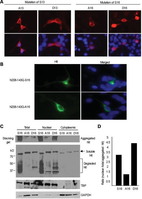
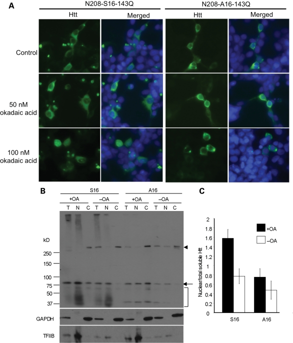
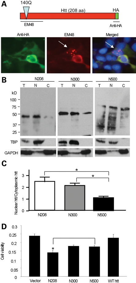
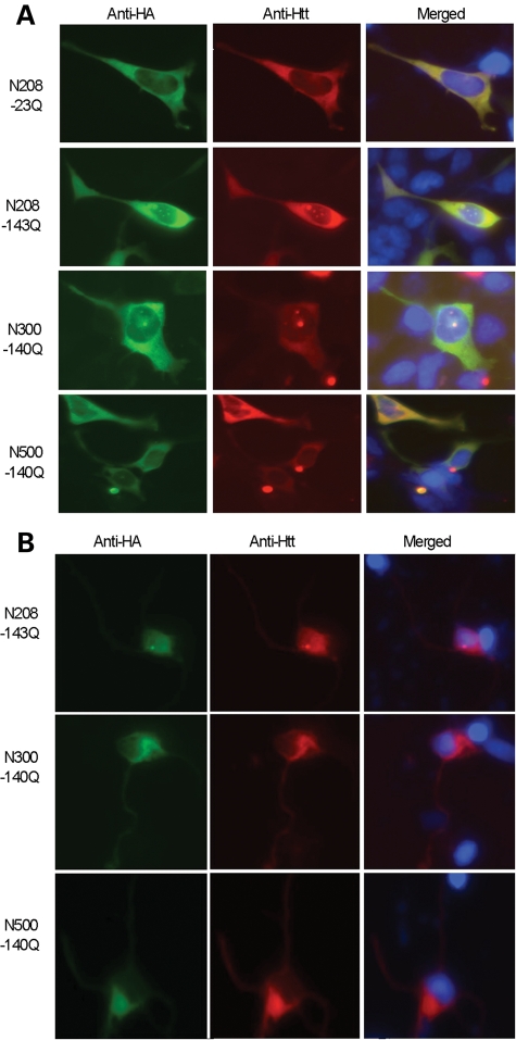
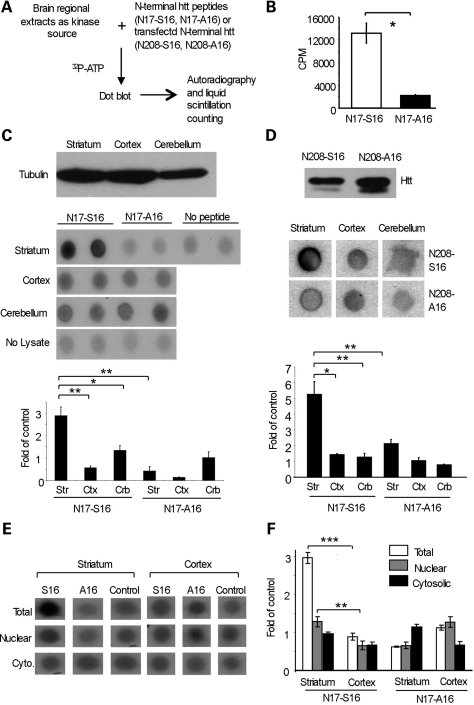
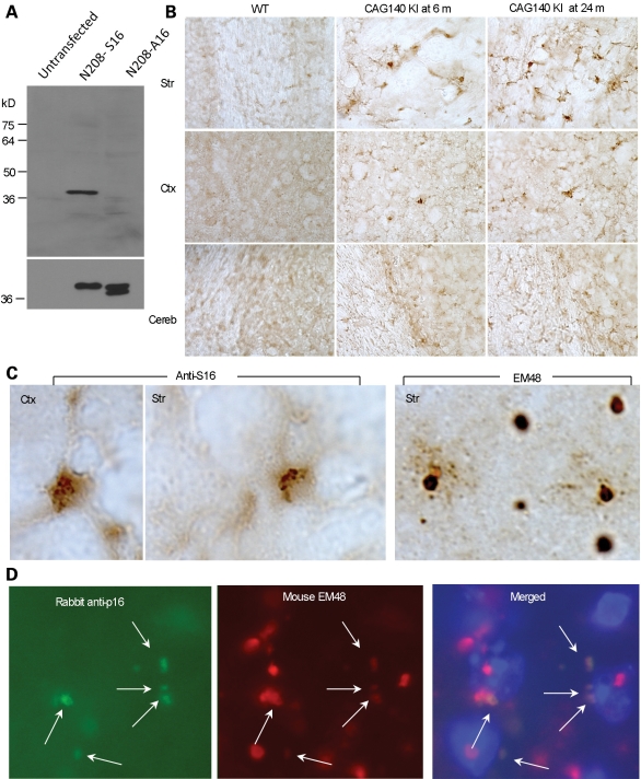
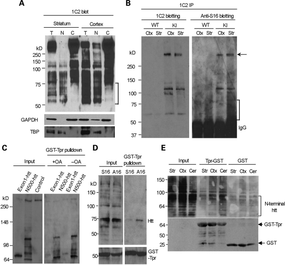
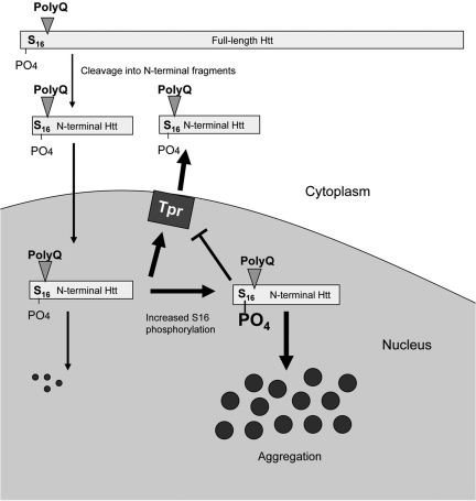
An expanded polyglutamine tract (>37 glutamines) in the N-terminal region of huntingtin (htt) causes htt to accumulate in the nucleus, leading to transcriptional dysregulation in Huntington disease (HD). In HD knock-in mice that express full-length mutant htt at the endogenous level, mutant htt preferentially accumulates in the nuclei of striatal neurons, which are affected most profoundly in HD. The mechanism underlying this preferential nuclear accumulation of mutant htt in striatal neurons remains unknown. Here, we report that serine 16 (S16) in htt is important for the generation of small N-terminal fragments that are able to accumulate in the nucleus and form aggregates. Phosphorylation of N-terminal S16 in htt promotes the nuclear accumulation of small N-terminal fragments and reduces the interaction of N-terminal htt with the nuclear pore complex protein Tpr. Mouse brain striatal tissues show increased S16 phosphorylation and a decreased association between mutant N-terminal htt and Tpr. These findings provide mechanistic insight into the nuclear accumulation of mutant htt and the selective neuropathology of HD, revealing potential therapeutic targets for treating this disease.


Figures

Figure 1.
Figure 1.
S16 of htt promotes the nuclear aggregation of N-terminal mutant htt. (A) HEK293 cells were transfected with N-terminal htt (N208-23Q), with S13 substituted by alanine (A13) or aspartic acid (D13), or with S16 substituted by alanine (A16) or aspartic acid (D16). Mimicking phosphorylation on S16 (D16) led to more nuclear distribution of htt than on S13 (D13). (B) Transfection of N208-143Q-S16 or -A16 in cultured rat striatal neurons showing that A16 substitution reduces the nuclear accumulation and aggregation of N-terminal mutant htt. (C) Subcellular fractionation of HEK293 cells transfected with mutant N-terminal htt (N208-143Q) containing S16, A16 or D16. EM48 western blotting reveals aggregated htt in the stacking gel, as well as soluble htt (arrow) and its degraded products (bracket) in the resolving gel. The blot was also probed with the antibody to the nuclear protein TATA-box-binding protein (TBP) and the cytoplasmic protein GAPDH. (D) The relative level of aggregated htt in the nucleus was determined using densitometry to calculate the ratio of nuclear aggregated htt to total aggregated htt.
Figure 2.
Figure 2.
OA-induced phosphorylation increased the levels of nuclear N208-143Q containing S16. (A) EM48 immunostaining of transfected HEK293 cells expressing N208-143Q-S16 or N208-143Q-A16 after treatment with OA (50 and 100 nm for 15 min). The control is cells without OA treatment. (B) Representative western blot (n = 3) of nuclear and cytosolic fractions of transfected HEK293 cells treated with 100 nm OA for 4 h. Substitution of S16 by alanine (A16) eliminated this increased nuclear level. The arrowhead indicates full-length normal htt in HEK293 cells. The arrow indicates soluble transfected htt. The bracket indicates degraded htt fragments. (C) Densitometry analysis of the ratio of nuclear soluble htt to htt in total lysates.
Figure 3.
Figure 3.
Shorter N-terminal htt fragments are more prone to nuclear accumulation than longer N-terminal fragments. (A) Expression of N208-143Q with the HA epitope at its C-terminus in HEK293 cells. Double immunofluorescent staining reveals that proteolytic N-terminal fragments without the HA epitope form nuclear aggregates (arrow) that were only labeled by EM48. (B) N-terminal htt (N208, N300 or N500) of varying lengths (208, 300 or 500 amino acids) with 140-143Q was transfected into HEK293 cells. Subcellular fractionation followed by western blotting with antibodies to htt (EM48), the nuclear protein TBP and the cytoplasmic protein GAPDH. (C) The ratio of nuclear to cytoplasmic htt was calculated via densitometry of western blots (n = 3) to compare the amounts of nuclear accumulation of each N-terminal fragment (N208, N300, N500). (D) HEK293 cells were transfected with N208, N300 or N500 containing 140Q for 48 h. A wild-type N-terminal htt with 23Q (N208-23Q) served as a control. Cell lysates were used for an MTS assay to compare cell viability expressing htt fragments of different sizes. *P < 0.05.
Figure 4.
Figure 4.
Small N-terminal mutant htt fragments show increased nuclear accumulation in transfected cells. EM48 immunostaining of HEK293 cells (A) or primary rat striatal neurons (B) transfected with N-terminal mutant htt of different lengths (208, 300 and 500 amino acids) containing 140-143Q. Transfected htt was tagged with the HA epitope at its C-terminus. The nuclei were stained with Hoechst dye.
Figure 5.
Figure 5.
Mouse striatal tissue lysates phosphorylate S16 of N-terminal htt at a higher level than lysates from other brain regions. (A) Lysates of the striatum, cortex and cerebellum from wild-type mice were incubated with N-terminal htt peptides or transfected N-terminal htt. The in vitro phosphorylation with 32P-ATP was analyzed by dot blotting and quantified by liquid scintillation counting. (B) The N-terminal 17 amino acid peptide of htt containing S16, but not A16, was phosphorylated in vitro using the method described in (A). (C) Western blotting for γ-tubulin was used to verify equal amounts of lysates (upper panel). Dot blot analysis (middle panel) and liquid scintillation counting (lower panel) of the in vitro phosphorylation reactions showed that striatal lysates significantly phosphorylated S16 of htt to a greater extent than the lysates from the cortex or cerebellum. (D) N208-23Q with S16 or A16 was expressed in transfected HEK293 cells and immunoprecipitated with EM48. Western blotting with mEM48 verified the precipitated htt (upper panel). Dot blot analysis (middle panel) and liquid scintillation counting (lower panel) from three independent experiments show that the precipitated N208-S16 htt was significantly more phosphorylated by the striatal lysates than the lysates from the cortex or cerebellum. (E) Total, nuclear and cytosolic fractions from the mouse striatum and cortex were used as the source of kinase to phosphorylate N17-16 and N17-A16. Dot blot analysis shows that the striatal nuclear fraction produced a higher level of S16 phosphorylation than the cytosolic fraction. (F) Liquid scintillation counting also revealed a significantly higher level of phosphorylated S16 by the striatal nuclear lysate, though the fractionation decreased the activity compared with total cell lysates. Control is the c.p.m. in the absence of peptides. *P < 0.05; **P < 0.01; ***P < 0.001.
Figure 6.
Figure 6.
htt with phosphorylated S16 is more abundant in the striatal neurons and is partly colocalized with EM48-positive small aggregates at the perinuclear region. (A) Generation of a phospho-S16 (anti-S16)-specific antibody. The specificity of anti-S16 for phosphorylated S16 was verified by western blotting of lysates from HEK293 cells expressing either N208-23Q-S16 or N208-23Q-A16 (upper panel). The same blot was also probed with the mEM48 antibody (lower panel). (B) Immunohistochemistry with anti-S16 was used to compare the levels of S16-phosphorylated htt in the striatum (Str), cortex (Ctx) and cerebellum (Cereb) of full-length htt (CAG140) KI mice at 6 or 24 months of age. S16-phosphorylated htt in the striatum increased with age and was more abundant than in the cortex. (C) Anti-S16 labeled diffuse htt and small aggregates, whereas EM48 intensively labeled large nuclear htt inclusions in the striatum of a CAG140 KI mouse. (D) Double immunostaining of the striatum of a CAG140 KI mouse with anti-S16 (green) and mEM48 (red) revealed that S16-phosphorylated htt was localized to mEM48-positive small perinuclear aggregates (arrows) in the perinuclear region. Hoechst dye was used to stain the nucleus.
Figure 7.
Figure 7.
N-terminal mutant htt with phosphorylated S16 is enriched in the nuclear fraction of striatal neurons and shows decreased binding to Tpr. (A) 1C2 western blotting revealing that N-terminal mutant htt fragments <75 kDa (marked by the bracket) are present only in the total and nuclear fractions, but not in the cytosolic fractions, of the mouse striatum and cortex. The same blot was also probed with antibodies to the cytoplasmic protein GAPDH and the nuclear protein TBP. (B) Mutant htt in CAG140 KI mouse striatum and cortex was immunoprecipitated by 1C2. Western blots with anti-S16 and 1C2 showed more abundant N-terminal fragments (marked by the bracket) in the striatum that were labeled by anti-S16. Densitometry analysis (lower panel) of the relative percentage of phosphorylated full-length htt by measuring the intensity of htt labeled by anti-S16 relative to htt labeled by 1C2. (C) HEK293 cells were cotransfected with GST-Tpr and exon1-150Q or N500-htt-140Q htt. More N500-htt-140Q and its degraded products than exon1-150Q were precipitated by GST-Tpr. Note that very little full-length normal htt (arrow) was precipitated and that increased phosphorylation by OA treatment reduces the interaction of N-terminal htt with GST-Tpr. (D) HEK293 cells were cotransfected with GST-Tpr (bracket) and N208-143Q containing S16 or A16. A GST pulldown followed by western blotting with mEM48 showed that phosphorylation elimination by A16 substitution increased the association of htt with Tpr. (E) Striatal, cortical and cerebellar lysates were prepared from CAG140 KI mice and incubated with GST-Tpr or GST alone. Western blotting of the GST pulldown samples with 1C2 showed that the binding of N-terminal htt fragments (marked by the bracket) to Tpr was reduced in the striatum. The blot was also probed with an antibody to GST to reveal GST and GST-Tpr used for the precipitation of mouse brain mutant htt.
Figure 8.
Figure 8.
Schematic diagram of increased S16 phosphorylation and nuclear accumulation of mutant htt. In the cytoplasm, full-length mutant htt is proteolytically degraded into small N-terminal htt fragments, which can enter the nucleus. Increased phosphorylation of S16 in N-terminal mutant htt enhances its aggregation and reduces its association with the nuclear pore complex protein Tpr, resulting in the accumulation of mutant htt in the nucleus. The size of N-terminal htt does not represent its actual proportion of full-length htt.

References

    1. DiFiglia M., Sapp E., Chase K.O., Davies S.W., Bates G.P., Vonsattel J.P., Aronin N. Aggregation of huntingtin in neuronal intranuclear inclusions and dystrophic neurites in brain. Science. 1997;277:1990–1993. doi:10.1126/science.277.5334.1990. - DOI - PubMed
    1. Gutekunst C.A., Li S.H., Yi H., Mulroy J.S., Kuemmerle S., Jones R., Rye D., Ferrante R.J., Hersch S.M., Li X.J. Nuclear and neuropil aggregates in Huntington's disease: relationship to neuropathology. J. Neurosci. 1999;19:2522–2534. - PMC - PubMed
    1. Orr H.T., Zoghbi H.Y. Trinucleotide repeat disorders. Annu. Rev. Neurosci. 2007;30:575–621. doi:10.1146/annurev.neuro.29.051605.113042. - DOI - PubMed
    1. Van Raamsdonk J.M., Murphy Z., Slow E.J., Leavitt B.R., Hayden M.R. Selective degeneration and nuclear localization of mutant huntingtin in the YAC128 mouse model of Huntington disease. Hum. Mol. Genet. 2005;14:3823–3835. doi:10.1093/hmg/ddi407. - DOI - PubMed
    1. Landles C., Bates G.P. Huntingtin and the molecular pathogenesis of Huntington's disease. Fourth in molecular medicine review series. EMBO Rep. 2004;5:958–963. doi:10.1038/sj.embor.7400250. - DOI - PMC - PubMed

Publication types

MeSH terms