. 2011 Feb 1;71(3):998-1008.
doi: 10.1158/0008-5472.CAN-09-3269. Epub 2011 Jan 18.

Smad4 inactivation promotes malignancy and drug resistance of colon cancer

Affiliations

Smad4 inactivation promotes malignancy and drug resistance of colon cancer

Panagiotis Papageorgis et al. Cancer Res. .

Abstract

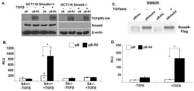
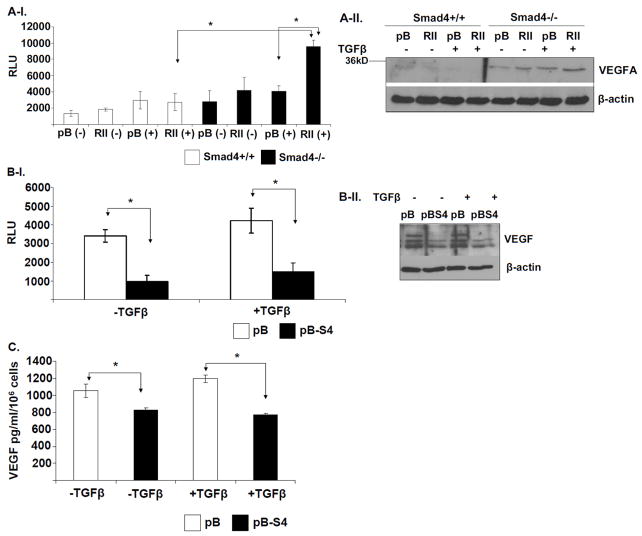
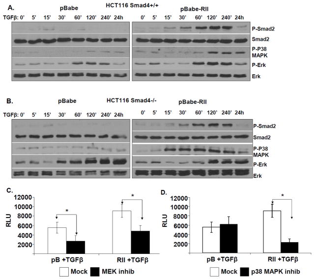
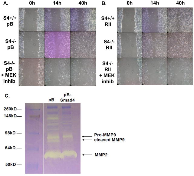
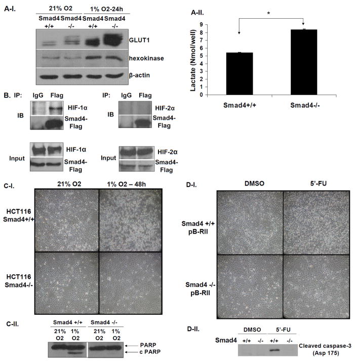
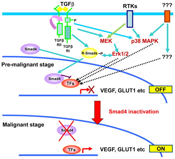
SMAD4 is localized to chromosome 18q21, a frequent site for loss of heterozygosity in advanced stage colon cancers. Although Smad4 is regarded as a signaling mediator of the TGFβ signaling pathway, its role as a major suppressor of colorectal cancer progression and the molecular events underlying this phenomenon remain elusive. Here, we describe the establishment and use of colon cancer cell line model systems to dissect the functional roles of TGFβ and Smad4 inactivation in the manifestation of a malignant phenotype. We found that loss of function of Smad4 and retention of intact TGFβ receptors could synergistically increase the levels of VEGF, a major proangiogenic factor. Pharmacologic inhibition studies suggest that overactivation of the TGFβ-induced MEK-Erk and p38-MAPK (mitogen-activated protein kinase) auxiliary pathways are involved in the induction of VEGF expression in SMAD4 null cells. Overall, SMAD4 deficiency was responsible for the enhanced migration of colon cancer cells with a corresponding increase in matrix metalloprotease 9 enhanced hypoxia-induced GLUT1 expression, increased aerobic glycolysis, and resistance to 5'-fluoruracil-mediated apoptosis. Interestingly, Smad4 specifically interacts with hypoxia-inducible factor (HIF) 1α under hypoxic conditions providing a molecular basis for the differential regulation of target genes to suppress a malignant phenotype. In summary, our results define a molecular mechanism that explains how loss of the tumor suppressor Smad4 promotes colorectal cancer progression. These findings are also consistent with targeting TGFβ-induced auxiliary pathways, such as MEK-ERK, and p38-MAPK and the glycolytic cascade, in SMAD4-deficient tumors as attractive strategies for therapeutic intervention.


Figures

Figure 1
Figure 1. Establishment and verification of HCT116 and SW620 colon cancer model cell lines
A. Western blotting of total cell lysates isolated from HCT116 SMAD4+/+ pBabe and pBabe-TGFβRII-HA as well as the isogenic SMAD4−/− pBabe and pBabe-TGFβRII-HA cells for detection of exogenously expressed TGFβRII-HA. B. SBE4-Luc luciferase reporter assay in the HCT116 cell lines. Cells were serum-starved overnight and treated with 5ng/ml TGFβ for 5h before lysis. C. Western blotting analysis of total cell lysates isolated from the stable SW620-pB and isogenic SW620-pBSMAD4 cells for detection of exogenously expressed Smad4-Flag. D. SBE4-Luc luciferase reporter assays in SW620 cells. Cells were serum-starved overnight and treated with 5ng/ml TGFβ for 5h before lysis.
Figure 2
Figure 2. Smad4 suppresses VEGF expression in colon cancer cells
A-I. Four groups of the indicated engineered HCT116 cell lines were either mock- treated or treated with 5ng/ml TGFβ for 24h. SBE4-Luc luciferase reporter assays were performed in the Mock- or TGFβ-treated HCT116 SMAD4+/+ (WT) pBabe and pBabe-TGFβRII-HA cells, mock- or TGFβ-treated SMAD4−/− (S4-) pBabe and pBabe-TGFβRII-HA cells. A-II. Western blotting was used to detect VEGF protein levels in corresponding total cell lysates. B-I. SW620-pB and isogenic SW620-pBSMAD4 cells were serum-starved overnight and treated with mock or 5ng/ml TGFβ for 24h. SBE4-Luc luciferase reporter assay was performed in the corresponding SW620 cells. Samples were measured in triplicates and the experiment was independently performed three times. II. Western blotting of total cell lysates was used to detect VEGF protein levels in the same four samples as indicated. C. Smad4 expression suppresses VEGF secretion from the SW620 cells. SW620-pB and SW620pBSmad4 cells were cultured in serum-free DMEM medium for 24h in the absence (-) or presence (+) of 5ng/ml TGFβ. Secreted VEGF was quantified by ELISA assay for VEGF in the conditioned media collected from each cell line. Results are presented as the average of triplicate measurements.
Figure 3
Figure 3. Involvement of MEK-Erk and p38MAPK pathways in the upregulation of VEGF in SMAD4 defective cells
A. HCT116 SMAD4+/+ pBabe and pBabe-TGFβRII-HA as well as B. SMAD4−/− pBabe and pBabe-TGFβRII-HA cells were serum-starved overnight and then treated with 2 ng/ml TGFβ for the indicated time periods. Western blotting of total cell lysates was used to monitor the kinetics of Smad2, Erk and p38MAPK phosphorylation. C & D. HCT116 SMAD4−/− pBabe and pBabe-RII-HA cells were cultured in serum-free medium and transfected with VEGF-luc and Renilla plasmids for 16h. Cells were then treated either with mock (DMSO) or with pharmacological inhibitors against MEK kinase (PD98059-20μM) (C) or p38 MAPK (SB203580–20μM) (D) for 30 min prior to mock- or TGFβ treatment (5 ng/ml) for an additional 24h. The samples were measured in triplicates and the experiment was independently performed three times.
Figure 4
Figure 4. Smad4 suppresses colon cancer cell migration and MMP9 activity
HCT116 SMAD4+/+ pBabe and SMAD4−/− pBabe (A) as well as pBabe-TGFβRII-HA and pBabe-TGFβRII-HA cells (B) were grown to confluency and then a cell-free area was introduced using a sterile Q-tip. Cells were either mock-treated or treated with 20μM MEK kinase inhibitor (PD98059) 30 minutes prior to introduction of the cell scratch. The ability of the cells to migrate into the cell-free area was monitored over time. Images show representative examples of three independent experiments. C. SW620-pB and SW620-pBSMAD4 cells were cultured in serum-free medium for 36h. Conditioned medium was collected and concentrated by centrifugation. Equal protein-containing samples were analyzed by zymogram gel assays. The gelatinolytic activities of MMP2, pro-MMP9 and cleaved MMP9 were detected by coommassie blue staining as clear bands on the gel at molecular weights corresponding to 60kD, 97kD and 85kD, respectively.
Figure 5
Figure 5. SMAD4 deficiency correlates with increased GLUT1 levels and resistance to hypoxia-induced cell death and 5′-fluorouracil treatment
A-I. Loss of SMAD4 increases GLUT1 protein levels. Western blotting for detection of GLUT1 protein levels in protein lysates isolated from HCT116 SMAD4+/+ and SMAD4−/− grown under normoxic (21% O2) or hypoxic (1% O2) conditions for 24h. A-II. Lactate secretion from HCT116 SMAD4+/+ and SMAD4−/− cells growing under normoxic conditions. B. Smad4 physically interacts with HIF1α but not with HIF2α under hypoxic conditions. HCT116 SMAD4+/+ cells were transiently co-transfected with PRK5-SMAD4-Flag and pCDNA3-HIF1αAA vectors or PRK5-SMAD4-Flag and pCDNA3-HIF2αAA vectors, respectively, for 16h and cultured under hypoxic conditions for an additional 5h. Total cell lysates were immunoprecipitated with mouse IgG antibody (mock) or mouse anti-Flag antibody and immunoprecipitates were analyzed by Western blotting to detect either HIF1α or HIF2α. C. Representative examples of light microscopy images (C-I) and Western blotting for detection of the cleaved PARP (C-II) in HCT116 SMAD4+/+ and SMAD4−/− grown in normoxic (21% O2) or hypoxic conditions (1% O2). D. Representative examples of light microscopy images (D-I) and Western blotting for detection of the cleaved caspase-3 (Asp 175) (D-II) from total cell lysates of HCT116 SMAD4+/+ and SMAD4−/− cultures which were either treated with either mock (DMSO) or 5′-fluorouracil (5′-FU) (1μg/ml) for 72h.
Figure 6
Figure 6. SMAD4 inactivation promotes transition to malignancy in colon cancer
Transition of pre-malignant colon cancer cells to malignancy is blocked by functional Smad4 due to inhibition of transcription factors (TFs) such as the HIF1α or other molecular events that are activated downstream of oncogenic signaling pathways and cross-talking TGFβ signaling events involved in promoting malignant properties. The inactivation of Smad4 during colon cancer progression removes the block in transition from the pre-malignant to the malignant stage by allowing accumulation of factors such as GLUT1 and VEGF.

References

    1. Jemal A, Siegel R, Ward E, Hao Y, Xu J, Thun MJ. Cancer statistics, 2009. CA Cancer J Clin. 2009;59:225–49. - PubMed
    1. Fearon ER, Vogelstein B. A genetic model for colorectal tumorigenesis. Cell. 1990;61:759–67. - PubMed
    1. Kinzler KW, Vogelstein B. Cancer-susceptibility genes. Gatekeepers and caretakers. Nature. 1997;386:761–63. - PubMed
    1. Thiagalingam S, Cheng K-h, Foy RL, Lee HJ, Chinnappan D, Ponte JF. TGF-beta and its Smad connection to cancer. Current Genomics. 2002;3:449–76.
    1. Massague J. TGF-beta signal transduction. Annu Rev Biochem. 1998;67:753–91. - PubMed

Publication types

MeSH terms

Substances