

Targeted signal-amplifying enzymes enhance MRI of EGFR expression in an orthotopic model of human glioma
- PMID: 21245103
- PMCID: PMC3059397
- DOI: 10.1158/0008-5472.CAN-10-1139
Targeted signal-amplifying enzymes enhance MRI of EGFR expression in an orthotopic model of human glioma
Abstract
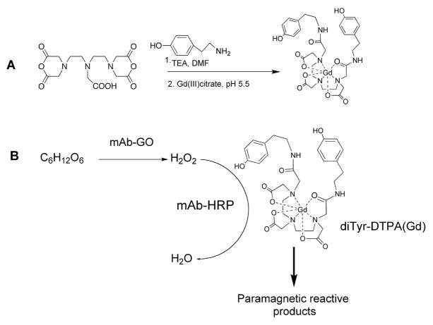
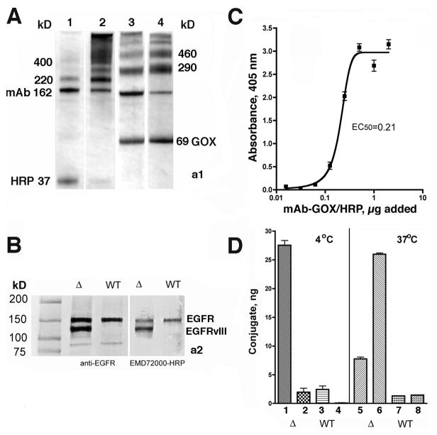
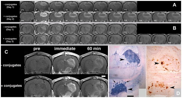
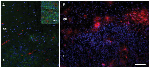
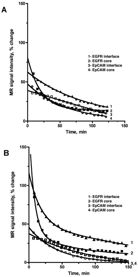
Epidermal growth factor receptor (EGFR) imaging in brain tumors is essential to visualize overexpression of EGFRvIII variants as a signature of highly aggressive gliomas and to identify patients that would benefit from anti-EGFR therapy. Seeking imaging improvements, we tested a novel pretargeting approach that relies on initial administration of enzyme-linked anti-EGFR monoclonal antibodies (mAb; EMD72000) followed by administration of a low-molecular-weight paramagnetic molecule (diTyr-GdDTPA) retained at the site of EGFR mAb accumulation. We hypothesized that diTyr-GdDTPA would become enzyme activated and retained on cells due to binding to tissue proteins. In support of this hypothesis, mAb-enzyme conjugates reacted with both membrane-isolated wild-type (wt) EGFR and EGFRvIII, but they bound primarily to EGFRvIII-expressing cells and not to EGFRwt-expressing cells. In vivo analysis of magnetic resonance (MR) tumor signal revealed differences in MR signal decay following diTyr-GdDTPA substrate administration. These differences were significant in that they suggested differences in substrate elimination from the tissue which relied on the specificity of the initial mAb binding: a biexponential signal decay was observed in tumors only upon preinjection with EGFR-targeted conjugates. Endpoint MRI in this setting revealed detailed images of tumors which correlated with immunohistochemical detection of EGFR expression. Together, our findings suggest an improved method to identify EGFRvIII-expressing gliomas in vivo that are best suited for treatment with therapeutic EGFR antibodies.
©2011 AACR.
Figures
Similar articles
-
MR signal amplification for imaging of the mutant EGF receptor in orthotopic human glioma model.Mol Imaging Biol. 2013 Dec;15(6):675-84. doi: 10.1007/s11307-013-0653-8. Mol Imaging Biol. 2013. PMID: 23733229 Free PMC article.
-
Nimotuzumab enhances temozolomide-induced growth suppression of glioma cells expressing mutant EGFR in vivo.Cancer Med. 2016 Mar;5(3):486-99. doi: 10.1002/cam4.614. Epub 2016 Jan 18. Cancer Med. 2016. PMID: 26778701 Free PMC article.
-
Targeted delivery of methotrexate to epidermal growth factor receptor-positive brain tumors by means of cetuximab (IMC-C225) dendrimer bioconjugates.Mol Cancer Ther. 2006 Jan;5(1):52-9. doi: 10.1158/1535-7163.MCT-05-0325. Mol Cancer Ther. 2006. PMID: 16432162
-
[177Lu]-Labeled 2-(4-isothiocyanatobenzyl)-6-methyldiethylene-triaminepentaacetic acid (1B4M-DTPA) conjugated monoclonal antibody L8A4 against epidermal growth factor receptor variant III (EGFRvIII).2010 Dec 24 [updated 2011 Feb 17]. In: Molecular Imaging and Contrast Agent Database (MICAD) [Internet]. Bethesda (MD): National Center for Biotechnology Information (US); 2004–2013. 2010 Dec 24 [updated 2011 Feb 17]. In: Molecular Imaging and Contrast Agent Database (MICAD) [Internet]. Bethesda (MD): National Center for Biotechnology Information (US); 2004–2013. PMID: 21348053 Free Books & Documents. Review.
-
[177Lu]-Labeled [(R)-2-amino-3-(4-isothiocyanatophenyl)propyl]-trans-(S,S)-cyclohexane-1,2-diamine-pentaacetic acid (CHX-A″-DTPA) conjugated monoclonal antibody L8A4 against epidermal growth factor receptor variant III (EGFRvIII).2010 Dec 24 [updated 2011 Feb 17]. In: Molecular Imaging and Contrast Agent Database (MICAD) [Internet]. Bethesda (MD): National Center for Biotechnology Information (US); 2004–2013. 2010 Dec 24 [updated 2011 Feb 17]. In: Molecular Imaging and Contrast Agent Database (MICAD) [Internet]. Bethesda (MD): National Center for Biotechnology Information (US); 2004–2013. PMID: 21348055 Free Books & Documents. Review.
Cited by
-
A novel paramagnetic substrate for detecting myeloperoxidase activity in vivo.Mol Imaging. 2012 Sep-Oct;11(5):433-43. Mol Imaging. 2012. PMID: 22954188 Free PMC article.
-
Molecular Magnetic Resonance Imaging of Tumors with a PTPµ Targeted Contrast Agent.Transl Oncol. 2013 Jun 1;6(3):329-37. doi: 10.1593/tlo.12490. Print 2013 Jun. Transl Oncol. 2013. PMID: 23730413 Free PMC article.
-
Tumor Detection at 3 Tesla with an Activatable Cell Penetrating Peptide Dendrimer (ACPPD-Gd), a T1 Magnetic Resonance (MR) Molecular Imaging Agent.PLoS One. 2015 Sep 3;10(9):e0137104. doi: 10.1371/journal.pone.0137104. eCollection 2015. PLoS One. 2015. PMID: 26336058 Free PMC article.
-
MR signal amplification for imaging of the mutant EGF receptor in orthotopic human glioma model.Mol Imaging Biol. 2013 Dec;15(6):675-84. doi: 10.1007/s11307-013-0653-8. Mol Imaging Biol. 2013. PMID: 23733229 Free PMC article.
-
A novel dual-labeled small peptide as a multimodal imaging agent for targeting wild-type EGFR in tumors.PLoS One. 2022 Feb 4;17(2):e0263474. doi: 10.1371/journal.pone.0263474. eCollection 2022. PLoS One. 2022. PMID: 35120180 Free PMC article.
References
-
- Bigner SH, Humphrey PA, Wong AJ, et al. Characterization of the epidermal growth factor receptor in human glioma cell lines and xenografts. Cancer Res. 1990;50:8017–22. - PubMed
-
- Schwechheimer K, Huang S, Cavenee WK. EGFR gene amplification--rearrangement in human glioblastomas. Int J Cancer. 1995;62:145–8. - PubMed
-
- Schlessinger J. Ligand-induced, receptor-mediated dimerization and activation of EGF receptor. Cell. 2002;110:669–72. - PubMed
-
- Shinojima N, Tada K, Shiraishi S, et al. Prognostic value of epidermal growth factor receptor in patients with glioblastoma multiforme. Cancer Res. 2003;63:6962–70. - PubMed
Publication types
MeSH terms
Substances
Grants and funding
LinkOut - more resources
Full Text Sources
Medical
Research Materials
Miscellaneous
