. 2011 Mar 18;286(11):9856-64.
doi: 10.1074/jbc.M110.196790. Epub 2011 Jan 18.

NAD+-dependent SIRT1 deacetylase participates in epigenetic reprogramming during endotoxin tolerance

Affiliations

NAD+-dependent SIRT1 deacetylase participates in epigenetic reprogramming during endotoxin tolerance

Tie Fu Liu et al. J Biol Chem. .

Abstract

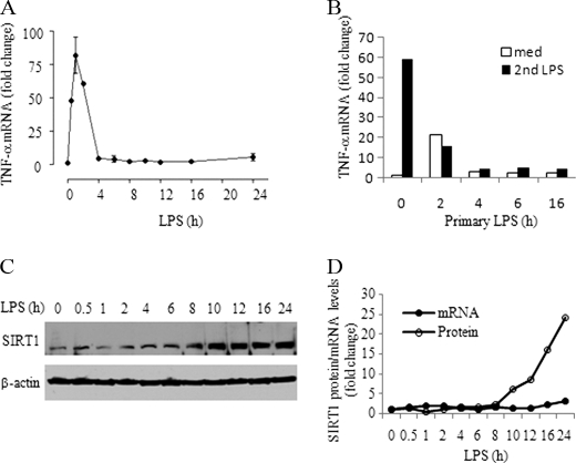
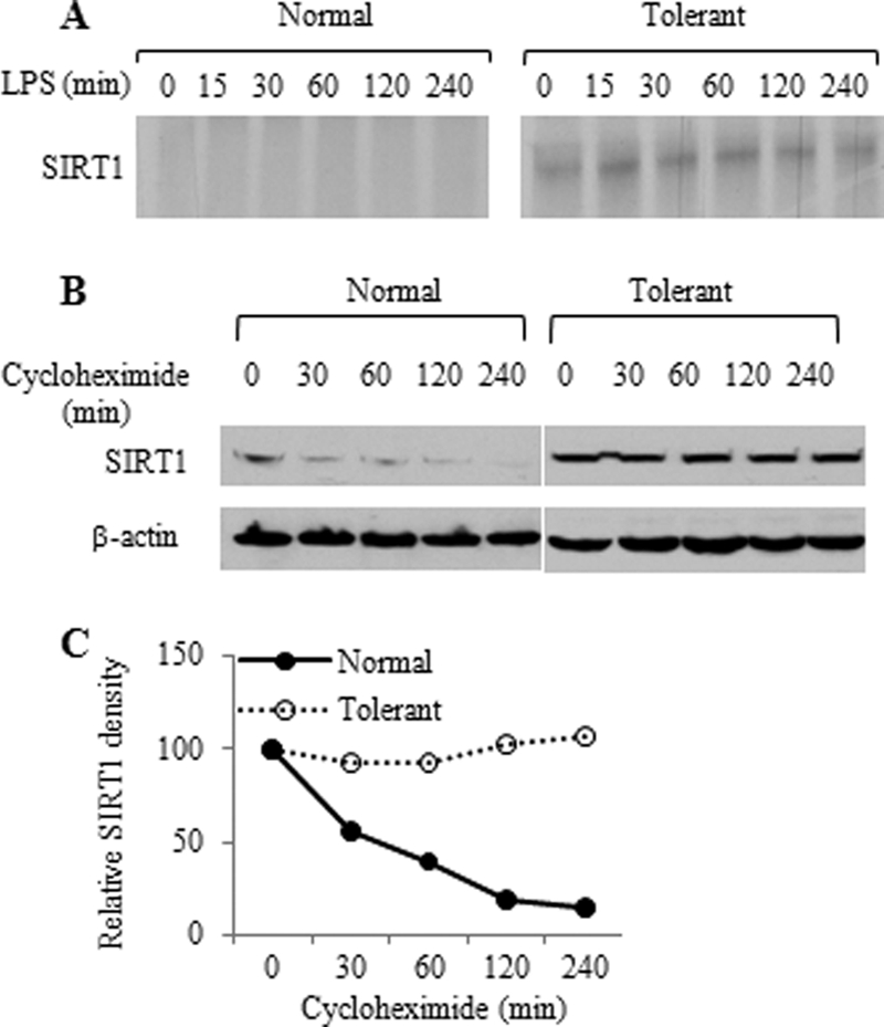
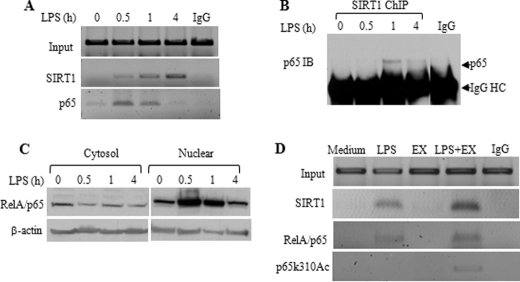
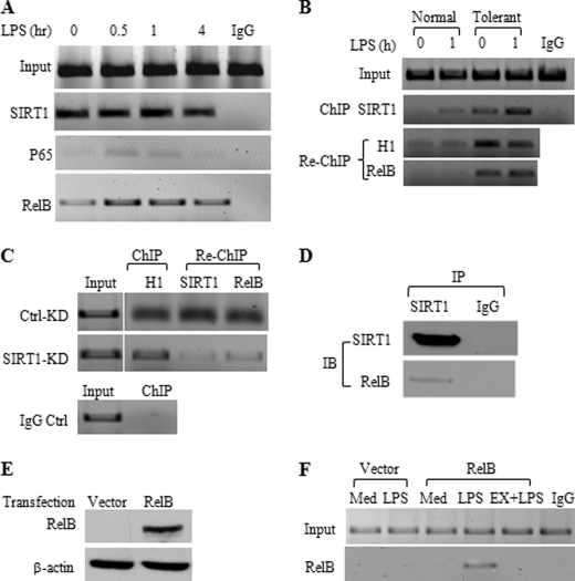
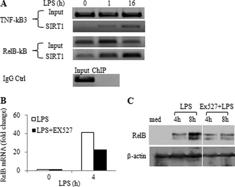
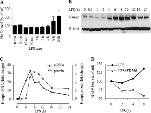
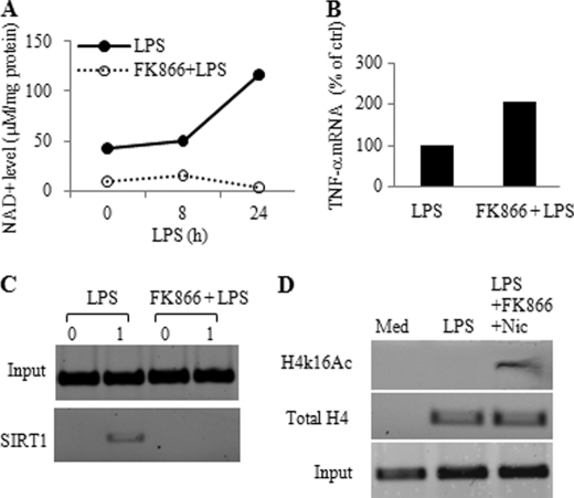
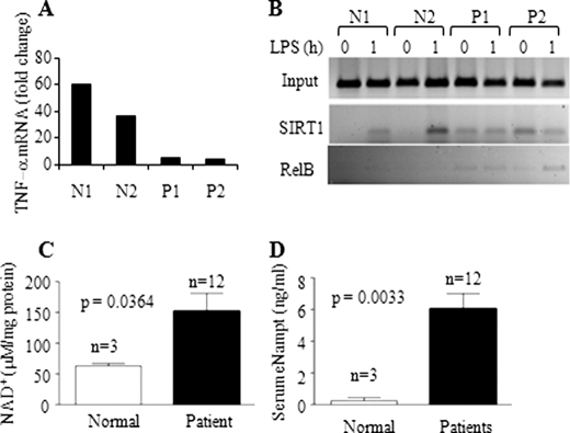
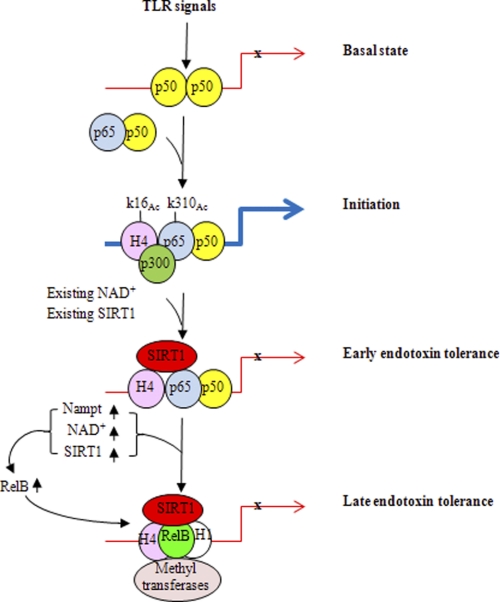
Gene-selective epigenetic reprogramming and shifts in cellular bioenergetics develop when Toll-like receptors (TLR) recognize and respond to systemic life-threatening infections. Using a human monocyte cell model of endotoxin tolerance and human leukocytes from acute systemic inflammation with sepsis, we report that energy sensor sirtuin 1 (SIRT1) coordinates the epigenetic and bioenergy shifts. After TLR4 signaling, SIRT1 rapidly accumulated at the promoters of TNF-α and IL-1β, but not IκBα; SIRT1 promoter binding was dependent on its co-factor, NAD(+). During this initial process, SIRT1 deacetylated RelA/p65 lysine 310 and nucleosomal histone H4 lysine 16 to promote termination of NFκB-dependent transcription. SIRT1 then remained promoter bound and recruited de novo induced RelB, which directed assembly of the mature transcription repressor complex that generates endotoxin tolerance. SIRT1 also promoted de novo expression of RelB. During sustained endotoxin tolerance, nicotinamide phosphoribosyltransferase (Nampt), the rate-limiting enzyme for endogenous production of NAD(+), and SIRT1 expression increased. The elevation of SIRT1 required protein stabilization and enhanced translation. To support the coordination of bioenergetics in human sepsis, we observed elevated NAD(+) levels concomitant with SIRT1 and RelB accumulation at the TNF-α promoter of endotoxin tolerant sepsis blood leukocytes. We conclude that TLR4 stimulation and human sepsis activate pathways that couple NAD(+) and its sensor SIRT1 with epigenetic reprogramming.


Figures

FIGURE 1.
FIGURE 1.
SIRT1 increases during TLR4-induced endotoxin tolerance in THP1 cells. A, kinetics of TNF-α transcription during the course of endotoxin tolerance. TNF-α mRNA levels were measured using quantitative real-time RT-PCR. B, TNF-α gene transcription was repressed in endotoxin-tolerant cells in response to second LPS stimulation. THP-1 cells pretreated with 1 μg/ml LPS for indicated times were restimulated with LPS for another 1 h. C, SIRT1 protein expression during the course of endotoxin tolerance. Normal THP-1 cells were stimulated for the indicated times with 1 μg/ml LPS, and cell lysates were subjected to Western blot analysis for SIRT1 level. β-Actin serves as loading control. D, real-time PCR analysis of SIRT1 transcription and densitometry analysis of SIRT1 protein expression in C. med, medium.
FIGURE 2.
FIGURE 2.
THP1 cells translate and stabilize SIRT1 following TLR4 stimulation. A, pulse chase analysis of SIRT1. Normal and tolerant THP-1 cells were pulsed for 1 h with [35S]methionine and chased for indicated times. The radiolabeled SIRT1 protein was immunoprecipitated with SIRT1 antibody and resolved onto 9% SDS-PAGE. B, SIRT1 protein degradation. Normal and tolerant cells were pretreated with 1 μm cycloheximide for 15 min followed by incubation with LPS for the indicated times. SIRT1 protein level in cell lysates was analyzed by Western blot analysis. C, densitometry analysis of the Western blot data shown in B.
FIGURE 3.
FIGURE 3.
SIRT1 represses TLR4-induced TNF-α transcription in normal and endotoxin-tolerant cells. A, effect of SIRT1-specific inhibitor Ex527 on TNF-α transcription. Normal THP-1 cells were pretreated with 1 μm Ex527 for 1 h followed by stimulation with 1 μg/ml LPS. TNF-α mRNA was quantified using real-time PCR. B, SIRT1 knockdown augments LPS-induced TNF-α transcription. Normal THP-1 cells were transfected with SIRT1-specific siRNA for 24 h as described under “Experimental Procedures.” Cells were then stimulated for 1 h with LPS. The insert shows Western blot analysis of SIRT1 protein levels. C, SIRT1 activator resveratrol inhibits LPS-induced TNF-α transcription. Normal THP-1 cells were pretreated for 1 h with 250 μm resveratrol followed by stimulation for 1 h with 1 μg/ml LPS. D, degradation of LPS-induced TNF-α mRNA. THP-1 cells were stimulated with 1 μg/ml LPS for 1 h in the presence or absence of Ex527 to induce maximal TNF-α mRNA. Cellular gene transcription was then inhibited by incubation of cells with 5 μg/ml of actinomycin D for indicated times. TNF-α mRNA levels are quantified using real-time PCR analysis and are presented as percentage of the maximum TNF-α mRNA. One of two similar experiment results is shown. SIRT1 inhibitor Ex527 (E) and SIRT1 knockdown (F) partially restore TNF-α transcription of tolerant THP-1 cells in response to second LPS stimulation. LPS-tolerant cells were treated as in A (Ex527) or as in B (SIRT1 knockdown) before LPS stimulation for 1 h. Data in A, B, C, E, and F are presented as percentage of control TNF-α mRNA and shown as mean ± S.E. Ctrl, control; KD, knockdown; Ex, Ex527; Res, resveratrol; ETOH, ethyl alcohol.
FIGURE 4.
FIGURE 4.
SIRT1 deacetylates RelA/p65 at lysine 310 at the TNF-α promoter in the initiation phase of TLR4 response. A, kinetics of SIRT1 and RelA/p65 association to the proximal TNF-α promoter. THP-1 cells were stimulated for the indicated times with 1 μg/ml LPS. Cell lysates were subjected to ChIP analysis using antibodies against SIRT1 or RelA/p65 as described under “Experimental Procedures.” B, SIRT1 interacts with RelA/p65 at promoters. THP-1 cells were stimulated for the indicated times, and chromatin-bound SIRT1 was immunoprecipitated using anti-SIRT1 antibody. SIRT1 immunoprecipitates were probed using anti-RelA/p65 antibody. C, LPS-induced dynamic changes of RelA/p65 protein. THP-1 cells were stimulated for indicated times, cytosol and nuclear extracts were prepared as described under “Experimental Procedures” and were subjected to Western blot analysis using anti-RelA/p65 antibody. D, SIRT1-specific inhibitor Ex527 accumulates acetylated RelA/p65 at lysine 310 (p65k310Ac) at proximal TNF-α promoter. THP-1 cells were cultured for 1 h in the absence or presence of 1 μg/ml LPS, 1 μm Ex527, or LPS plus Ex527, respectively. Cell lysates were subjected to ChIP assay using the indicated antibodies. IB, immunoblotting; EX, Ex527; HC, heavy chain.
FIGURE 5.
FIGURE 5.
SIRT1 accumulates with RelB and H1 at the TNF-α promoter. A, ChIP analysis of TNF-α promoter-bound SIRT1, RelA/p65, and RelB in tolerant cells. LPS-tolerant cells were restimulated for indicated times with 1 μg/ml LPS. Cell lysates were subjected to ChIP analysis using indicated antibodies as detailed under “Experimental Procedures.” B, SIRT1 interacts with H1 and RelB at the TNF-α promoter of tolerant cells. Normal and tolerant cells were stimulated for 1 h by LPS, SIRT1/H1, and RelB sequential ChIP analysis was performed as detailed under “Experiment Procedures.” C, H1/SIRT1 and RelB sequential ChIP analysis at the TNF-α promoter after SIRT1 knockdown. Cells were stimulated with LPS for 12 h followed by SIRT1 knockdown for 24 h. Chromatin was immunoprecipitated with H1 antibody and reimmunoprecipitated with SIRT1 or RelB antibody. D, SIRT1 interacts with RelB. THP-1 cells were stimulated for 24 h with 1 μg/ml LPS, nuclear extracts were immunoprecipitated with SIRT1 antibody. Immunoprecipitates were subjected to Western blot analysis using anti-RelB antibody. E, overexpression of RelB in THP-1 cells. Normal THP-1 cells were transfected for 48 h with 0.5 μg of either pcDNA3-HA vector plasmid DNA or HA-RelB plasmid DNA. Total cell lysates were subjected to Western blot analysis of RelB expression using anti-RelB antibody. F, SIRT1 facilitates RelB loading onto the TNF-α promoter. RelB-transfected THP-1 cells as detailed in E were stimulated for 1 h with LPS in the presence or absence of Ex527. ChIP analysis of TNF-α promoter bound RelB was performed as described under “Experimental Procedures.” IP, immunoprecipitation; IB, immunoblotting; Ctrl, control; KD, knockdown; Med, medium; EX, EX527.
FIGURE 6.
FIGURE 6.
SIRT1 regulates RelB expression. A, SIRT1 binds to RelB promoter. THP-1 cells were stimulated with 1 μg/ml of LPS for 0, 1, or 16 h, and chromatin was immunoprecipitated with SIRT1 antibody. The SIRT1-DNA complex was analyzed by standard PCR using TNF-α promoter-specific or RelB promoter-specific primers. IgG-immunoprecipitated samples served as negative control. B and C, inhibition of SIRT1 deacetylase activity decreases LPS-induced RelB gene transcription and protein expression during the development of endotoxin tolerance. THP-1 cells were pretreated for 1 h with 1 μm Ex527 followed by stimulation with 1 μg/ml of LPS for indicated times. RelB mRNA was quantified by real-time PCR and RelB protein levels were analyzed using Western blot. RelB mRNA levels are presented as fold changes relative to unstimulated control. Ctrl, control; med, medium.
FIGURE 7.
FIGURE 7.
Nampt expression and cellular NAD+ increase during TLR4-induced endotoxin tolerance. A, changes in cellular NAD+ levels during the course of endotoxin tolerance. Cells were cultured for different times in the presence of 1 μg/ml LPS. Intracellular NAD+ was extracted and evaluated using a commercial kit as detailed under “Experimental Procedures”. Results are reported as mean ± S.E. of 3 independent experiments. B: Western blot analysis of Nampt expression at the indicated times after 1 μg/ml of LPS stimulation. C, Real-time PCR analysis of Nampt transcription after LPS stimulation and densitometry analysis of Nampt protein levels in B. D, Nampt inhibitor FK866 inhibits LPS-induced NAD+ biosynthesis and depletes cellular NAD+. THP-1 cells were cultured for the indicated times with 1 μg/ml LPS or LPS plus 10 nm FK866. Intracellular NAD+ was extracted and analyzed as in A.
FIGURE 8.
FIGURE 8.
Cellular NAD+ is required for SIRT1 promoter binding and repression of TNF-α transcription. A, cellular NAD+ is diminished by the Nampt-specific inhibitor FK866. THP-1 cells were pretreated for 24 h with 10 nm FK866. Cells were washed and stimulated for indicated times with 1 μg/ml LPS in the presence of 10 nm FK866. B, depletion of cellular NAD+ by FK866 augments LPS-induced TNF-α transcription. THP-1 cells were pretreated for overnight with 10 nm FK866 followed by stimulation for 1 h with 1 μg/ml LPS. TNF-α mRNA was quantified using real-time PCR. C, depletion of cellular NAD+ inhibits SIRT1 binding to the TNF-α promoter. THP-1 cells were treated as in A. Cell lysates were subjected to SIRT1 ChIP assay at the TNF-α proximal promoter. D, depleting NAD+ by FK866 simultaneously with inhibiting SIRT1 activity enhances accumulation of acetylated histone H4 at lysine 16. Cells were treated for 16 h with LPS in the presence or absence of 10 nm FK866 and 1 mm nicotinamide followed by ChIP analysis with H4K16Ac antibody. Med, medium; Nic, nicotinamide.
FIGURE 9.
FIGURE 9.
Increases in cellular NAD+ and extracellular Nampt and accumulation of SIRT1 and RelB at TNF-α promoter in septic leukocytes. A, TNF-α transcription in blood leukocytes in response to LPS stimulation. Blood leukocytes were isolated from normal and septic subjects and stimulated with LPS for 1 h. RNA was isolated and analyzed for TNF-α mRNA by real-time PCR. B, ChIP analysis of TNF-α promoter-bound SIRT1 and RelB in normal and septic leukocytes. C, intracellular NAD+ levels in normal (n = 3) and septic blood leukocytes (n = 12). D, extracellular Nampt levels in normal (n = 3) and septic serum (n = 12). Data in C and D are shown as mean ± S.E. N, normal control; P, septic patient.
FIGURE 10.
FIGURE 10.
A model for SIRT1 in bridging bioenergetics and epigenetics during endotoxin tolerance and sepsis. TLR induces rapid promoter binding of constitutive SIRT1, which uses available nuclear NAD+ to support its promoter binding and deactivate RelA/p65 through lysine 310 deacetylation, thus limiting transcription of acute proinflammatory genes. SIRT1 then remains promoter bound and TLR-dependent responses increase the expression of Nampt, which sustains NAD+ elevation by recycling nicotinamide. Increases in Nampt are accompanied by elevations in SIRT1 by stabilizing the protein and enhancing translation. The combined availability of nuclear NAD+ and its effect on SIRT1 promoter accumulation stimulates RelB expression and supports recruitment of RelB to direct formation of locus-specific facultative heterochromatin to silence acute proinflammatory genes or its activating gene sets with distinct functions.

References

    1. Schreiber J., Jenner R. G., Murray H. L., Gerber G. K., Gifford D. K., Young R. A. (2006) Proc. Natl. Acad. Sci. U.S.A. 103, 5899–5904 - PMC - PubMed
    1. Medzhitov R., Horng T. (2009) Nat. Rev. Immunol. 9, 692–703 - PubMed
    1. McCall C. E., Yoza B., Liu T., El Gazzar M. (2010) J. Innate Immun. 2, 395–405 - PMC - PubMed
    1. Hotchkiss R. S., Karl I. E. (2003) N. Engl. J. Med. 348, 138–150 - PubMed
    1. McCall C. E., Yoza B. K. (2007) Am. J. Respir. Crit. Care Med. 175, 763–767 - PMC - PubMed

Publication types

MeSH terms