. 2011 Feb 1;108(5):2046-51.
doi: 10.1073/pnas.1014188108. Epub 2011 Jan 18.

LTβR signaling in dendritic cells induces a type I IFN response that is required for optimal clonal expansion of CD8+ T cells

Affiliations

LTβR signaling in dendritic cells induces a type I IFN response that is required for optimal clonal expansion of CD8+ T cells

Leslie Summers deLuca et al. Proc Natl Acad Sci U S A. .

Abstract

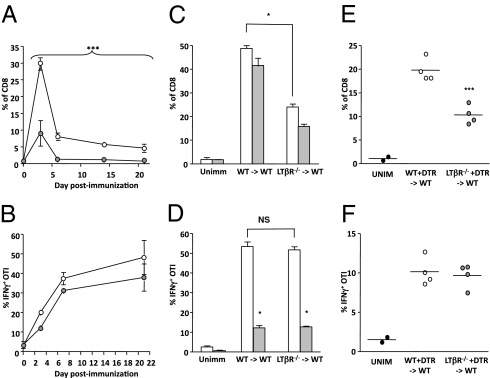
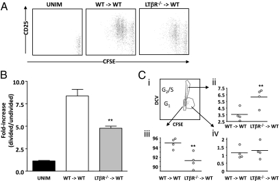
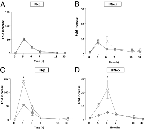
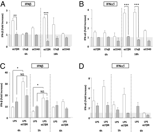
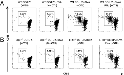
During an immune response, antigen-bearing dendritic cells (DCs) migrate to the local draining lymph node and present antigen to CD4(+) helper T cells. Antigen-activated CD4(+) T cells then up-regulate TNF superfamily members including CD40 ligand and lymphotoxin (LT)αβ. Although it is well-accepted that CD40 stimulation on DCs is required for DC licensing and cross-priming of CD8(+) T-cell responses, it is likely that other signals are integrated into a comprehensive DC activation program. Here we show that a cognate interaction between LTαβ on CD4(+) helper T cells and LTβ receptor on DCs results in unique signals that are necessary for optimal CD8(+) T-cell expansion via a type I IFN-dependent mechanism. In contrast, CD40 signaling appears to be more critical for CD8(+) T-cell IFNγ production. Therefore, different TNF family members provide integrative signals that shape the licensing potential of antigen-presenting DCs.


Conflict of interest statement

The authors declare no conflict of interest.

Figures

Fig. 1.
Fig. 1.
DC-derived LTβR and CD40 signals contribute distinctly to CD8+ T-cell cross-priming in vivo. (A and B) WT bm1xB6 F1 mice were given an A/T of responder CD45.1 or Thy1.1 OTI T cells, treated with either control human IgG (huIgG) or LTβR-Ig, and immunized the next day with OVA-loaded bm1 cells. At multiple days postimmunization, OTI expansion (A) and IFNγ production (B) were measured. Data are representative of six (A) or two (B) independent experiments (n = 3–6 per experiment). (C and D) WT→WT or LTβR−/−→WT chimeric mice were given an A/T of responder CD45.1 or Thy1.1 OTI T cells, treated with either control Ab or α-CD40L, and immunized with OVA-loaded bm1 cells, and OTI expansion (C) and IFNγ production (D) were measured at day 3. These experiments were performed four times with similar results, and results shown represent the average of four mice per group. NS, nonsignificant. (E and F) Mixed chimeric WT + CD11c-DTR→WT or LTβR−/− + CD11c-DTR→WT mice were given an A/T of responder CD45.1 OTI T cells, treated with diphtheria toxin, and immunized with OVA-loaded bm1 cells, and OTI expansion (E) and IFNγ production (F) were measured at day 3. Data are representative of two experiments (n = 4 per experiment). Empty circles, control Ig-treated mice; filled circles, LTβR-Ig–treated mice; white bars, control treated mice; gray bars, α-CD40L–treated mice. *P < 0.05, ***P < 0.0001 (two-way ANOVA for A).
Fig. 2.
Fig. 2.
LTβR signaling is required for normal CD8+ T-cell activation and cell-cycle completion. Chimeric mice (WT→WT or LTβR−/−→WT) were given an A/T of CFSE-labeled responder OTI T cells, and then were either left unimmunized or immunized 1 d later with OVA-loaded bm1 cells. At day 2 postimmunization, CFSE dilution and CD25 up-regulation were evaluated (A), and the extent of CD25 up-regulation on divided versus undivided OTI cells was measured (B). Data are representative of three independent experiments (n = 3–5 mice per experiment). **P < 0.01. (C) To assess cell cycling in the described in vivo experiment from A and B, CFSE-stained OTI T cells were gated and analyzed with DyeCycle Violet to measure cell-cycle status, with a representative FACS plot shown (i). Enumeration of OTI that had divided [CFSEmed (ii)], had “terminally divided” [CFSE (iii)], or had remained undivided [CFSEhigh (iv)] was performed. Data are representative of two experiments (in one case performed at day 2, and another at day 3, n = 4 per experiment). Note that in the presence of DyeCycle Violet, CFSE intensity appears different from without DyeCycle Violet (A versus C). **P < 0.01.
Fig. 3.
Fig. 3.
LTβR synergizes with TLR4 to maximize type I IFN production in BMDCs. WT (open circles) and LTβR−/− (gray circles) BMDCs were coincubated with OVA-specific CD4+ T cells (OTII) and LPS (A and B). In some cases, these cocultures were also supplemented with OVA323–339 (C and D). DCs were isolated at indicated time points and the expression of IFNβ (A and C) and IFNα5 (B and D) was measured by real-time RT-PCR. This experiment was performed three times with similar results. *P < 0.05.
Fig. 4.
Fig. 4.
LTβR signaling can mediate type I IFN production independently of TLR activation in BMDCs. BMDCs were stimulated with anti-LTβR, LTαβ, or anti-CD40 in the absence of any added LPS. Expression of IFNβ (A) and IFNα5 (B) was measured by real-time RT-PCR at 6 and 18 h poststimulation of WT (open bars) and LTβR−/− (gray bars) BMDCs. These experiments were performed at least three times with similar results. *P < 0.05, **P < 0.01, ***P < 0.001. BMDCs were also preincubated with LPS for 2 h, washed, and then stimulated with anti-LTβR. Expression of IFNβ (C) and IFNα5 (D) was measured by real-time RT-PCR at 4, 5, and 6 h poststimulation of WT (open bars) and LTβR−/− (gray bars) BMDCs. This experiment was performed three times with similar results. *P < 0.05 for data in (C).
Fig. 5.
Fig. 5.
LTβR−/− BMDCs do not support full OTI proliferation in vitro, and this can be rescued with type I IFN. WT (A) and LTβR−/− (B) BMDCs were preincubated with LPS and OVA protein for 18 h, washed, and then plated with CFSE-labeled OVA-specific CD8+ T cells (OTI) with or without OTII CD4+ T cells. Seventy-two hours later, OTI T cells were gated based on CD45.1 expression and/or CD8 expression, and then assessed for CD25 expression and CFSE dilution. In some cases, 25 U/mL of IFNα was added to the cultures at 48 h. The experiment is a representative example of three independent experiments.

References

    1. Bevan MJ. Helping the CD8(+) T-cell response. Nat Rev Immunol. 2004;4:595–602. - PubMed
    1. Hochweller K, Anderton SM. Kinetics of costimulatory molecule expression by T cells and dendritic cells during the induction of tolerance versus immunity in vivo. Eur J Immunol. 2005;35:1086–1096. - PubMed
    1. Summers-deLuca LE, et al. Expression of lymphotoxin-αβ on antigen-specific T cells is required for DC function. J Exp Med. 2007;204:1071–1081. - PMC - PubMed
    1. Bennett SR, et al. Help for cytotoxic-T-cell responses is mediated by CD40 signalling. Nature. 1998;393:478–480. - PubMed
    1. Schoenberger SP, Toes RE, van der Voort EI, Offringa R, Melief CJ. T-cell help for cytotoxic T lymphocytes is mediated by CD40-CD40L interactions. Nature. 1998;393:480–483. - PubMed

Publication types

Substances

LinkOut - more resources