. 2011 Feb 1;108(5):2034-9.
doi: 10.1073/pnas.1016229108. Epub 2011 Jan 18.

HRD1 and UBE2J1 target misfolded MHC class I heavy chains for endoplasmic reticulum-associated degradation

Affiliations

HRD1 and UBE2J1 target misfolded MHC class I heavy chains for endoplasmic reticulum-associated degradation

Marian L Burr et al. Proc Natl Acad Sci U S A. .

Abstract

The assembly of MHC class I molecules is governed by stringent endoplasmic reticulum (ER) quality control mechanisms. MHC class I heavy chains that fail to achieve their native conformation in complex with β2-microglobulin (β2m) and peptide are targeted for ER-associated degradation. This requires ubiquitination of the MHC class I heavy chain and its dislocation from the ER to the cytosol for proteasome-mediated degradation, although the cellular machinery involved in this process is unknown. Using an siRNA functional screen in β2m-depleted cells, we identify an essential role for the E3 ligase HRD1 (Synoviolin) together with the E2 ubiquitin-conjugating enzyme UBE2J1 in the ubiquitination and dislocation of misfolded MHC class I heavy chains. HRD1 is also required for the ubiquitination and degradation of the naturally occurring hemochromatosis-associated HFE-C282Y mutant, which is unable to bind β2m. In the absence of HRD1, misfolded HLA-B27 accumulated in cells with a normal MHC class I assembly pathway, and HRD1 depletion prevented the appearance of low levels of cytosolic unfolded MHC I heavy chains. HRD1 and UBE2J1 associate in a complex together with non-β2m bound MHC class I heavy chains, Derlin 1, and p97 and discriminate misfolded MHC class I from conformational MHC I-β2m-peptide heterotrimers. Together these data support a physiological role for HRD1 and UBE2J1 in the homeostatic regulation of MHC class I assembly and expression.


Conflict of interest statement

The authors declare no conflict of interest.

Figures

Fig. 1.
Fig. 1.
Ubiquitin E3 ligase HRD1 is required for the degradation of β2m-deficient MHC I. (A and B) Cytofluorometric analysis of GFP levels in HeLa GFP-HLA-A2 shβ2m cells on siRNA depletion of ER membrane E3 ligases. (A) HRD1 depletion significantly increases GFP signal. Z scores calculated from two independent experiments. Z score of 3.0 (dashed line) equates to P = 0.05 (Bonferroni corrected). Numbers on y axis refer to E3s tested (Table S1). (B) GFP signal in siRNA treated (black line) vs. mock (shaded) HeLa GFP-HLA-A2 shβ2m cells. (C) HRD1 depletion rescues endogenous MHC I in β2m-depleted HeLa cells. Immunoblot for HRD1 and MHC I on siRNA depletion of β2m with or without HRD1. (D–F) HRD1-C1A RING mutant has a dominant negative effect rescuing misfolded MHC I from degradation. (D) GFP levels in HeLa GFP-HLA-A2 shβ2m cells transfected with wild-type HRD1 (HRD1wt), HRD1-C1A, or vector control. Tomato red fluorescent protein (RFP) was cotransfected (10%) as a marker of HRD1 expression. (E and F) HRD1-C1A rescues endogenous MHC I in β2m-depleted cells. (E) Immunoblot analysis of MHC I, HRD1, and control calreticulin in siRNA β2m-depleted HeLa cells after transfection with HRD1wt, HRD1-C1A, or vector control. (F) Immunoblot for MHC I and control calnexin in 293T cells cotransfected with β2m shRNA and either HRD1wt, HRD1-C1A, or vector control. Molecular mass in kilodaltons is indicated on the left for immunoblots throughout.
Fig. 2.
Fig. 2.
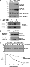
Misfolded MHC I HCs interact with HRD1 and are retained in the ER in its absence. (A) HRD1 associates with misfolded MHC I. β2m or mock siRNA-depleted HeLa cells were treated with or without IFN-γ 200 U/mL for 12 h and MG132 50 μM for 5 h, before lysis in 1% Digitonin, MHC I immunoprecipitation with HC10, and immunoblot for HRD1 and MHC I (3B10.7). (B) HRD1 depletion prevents dislocation of misfolded MHC I from the membrane to the cytosol. β2m-depleted HeLa cells were fractionated into membrane (M) and soluble (S) fractions. MHC I HCs were immunoprecipitated (HC10) from each fraction and visualized by immunoblot (3B10.7). In the presence of MG132 (40 μM for 4 h), MHC I HC is visible in the soluble fraction on β2m depletion but is retained in the membrane on additional HRD1 depletion. (C) HRD1 depletion prevents dislocation of MHC I HC in wild-type HeLa cells. HeLa cells depleted of HRD1 or gp78 were treated as in B. Low levels of MHC I HC are visible in the soluble fraction after long exposure of immunoblots but are not detected on HRD1 depletion. (D) HeLa cells depleted of β2m alone or codepleted of β2m and HRD1 were radiolabeled with 35S and MHC I HCs immunoprecipitated (HC10) from detergent lysates at the indicated chase times.
Fig. 3.
Fig. 3.
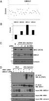
E2 ubiquitin-conjugating enzyme UBE2J1 is required for ubiquitination and degradation of β2m-deficient MHC I. (A and B) Cytofluorometric analysis of GFP levels in HeLa GFP-HLA-A2 shβ2m cells on siRNA depletion of E2 ubiquitin-conjugating enzymes. Z scores calculated from two independent experiments. A Z score of 3.2 (dashed line) equates to P = 0.05 (Bonferroni corrected). Numbers on y axis refer to E2s tested (Table S2). (B) Z scores of ER membrane-associated E2 enzymes. Only UBE2J1 depletion significantly rescues GFP signal. (C) UBE2J1 depletion rescues endogenous MHC I in β2m-depleted HeLa cells. Immunoblot of MHC I, UBE2J1, and control calnexin on siRNA depletion of β2m and E2 enzymes UBE2J1(J1), UBE2J2(J2), and UBE2G2(G2). (D) HRD1 and UBE2J1 depletion prevents ubiquitination of MHC I. β2m siRNA and mock-depleted HeLa cells were depleted of HRD1 with or without UBE2J1. After incubation with MG132 50 μM, lysates were immunoprecipitated with HC10 (MHC I HC) and probed for polyubiquitin (P4D1) and MHC I (3B10.7). Lysates (10%) were directly probed for HRD1, UBE2J1, and calnexin.
Fig. 4.
Fig. 4.
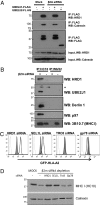
HRD1 and UBE2J1 are found in a complex with unfolded MHC I, p97, and Derlin 1. (A) UBE2J1 associates with HRD1 and calnexin. β2m or mock siRNA-depleted HeLa cells were transfected with UBE2J1-FLAG or UBE2J2-FLAG. After incubation with MG132 50 μM, 1% Digitonin lysates were immunoprecipitated with anti-FLAG antibody (M2) and probed for HRD1 and calnexin. (B) β2m or mock siRNA-depleted HeLa cells were pretreated with IFN-γ for 12 h and MG132 50 μM for 5 h, before lysis in 1% Digitonin. Each sample was split and immunoprecipitated with either HC10 or W6/32 before immunoblot for HRD1, UBE2J1, Derlin 1, p97, and 3B10.7. *Secondary antibody cross-reacting with antibody HC. (C and D) Depletion of SEL1L protects misfolded MHC I from degradation. (C) GFP levels in siRNA β2m-depleted HeLa GFP-HLA-A2 cells on siRNA depletion (black line) of SEL1L, HRD1, gp78, or TRC8, vs. mock (shaded). (D) Immunoblot analysis of endogenous MHC I in samples treated as in C.
Fig. 5.
Fig. 5.
HRD1 is required for the degradation of HFE-C282Y and HLA-B27 HC. (A and B) HRD1 depletion prevents ubiquitination of FLAG-HFE-C282Y and inhibits its degradation. (A) HRD1 or mock siRNA-depleted FLAG-HFE-C282Y–expressing HeLa cells were 35S radiolabeled and FLAG-HFE-C282Y immunoprecipitated (anti-FLAG) from lysates taken at the indicated chase times. (B) HRD1 or mock siRNA-depleted HeLa FLAG-HFE-C282Y cells were treated with or without MG132 50 μM before Triton X-100 lysis, immunoprecipitation of FLAG-HFE-C282Y (anti-FLAG), and immunoblot for polyubiquitin (P4D1). Lysates (10%) were directly immunoblotted for FLAG-HFE-C282Y and calnexin control. (C) HRD1 interacts with HFE-C282Y. HeLa FLAG-HFE-C282Y cells and control HeLa cells were treated with or without MG-132 50 μM before lysis in 1% Digitonin, immunoprecipation of FLAG-HFE-C282Y, and immunoblot for HRD1. (D and E) HRD1 depletion leads to accumulation of HLA-B27 HC dimers. (D) Lysates from HRD1 or mock siRNA-depleted HeLa cells expressing HLA-B27, HLA-B14, or HLA-A34 were separated by nonreducing or reducing SDS/PAGE and immunoblotted with HC10. (E) HRD1 or mock siRNA-depleted HeLa and HeLa HLA-B27 cells were treated with or without MG-132 50 μM. Triton X-100 lysates were immunoprecipitated sequentially with HC10 then W6/32, separated by nonreducing or reducing SDS/PAGE, and immunoblotted with 3B10.7.

References

    1. Wearsch PA, Cresswell P. The quality control of MHC class I peptide loading. Curr Opin Cell Biol. 2008;20:624–631. - PMC - PubMed
    1. Chapman DC, Williams DB. ER quality control in the biogenesis of MHC class I molecules. Semin Cell Dev Biol. 2010;21:512–519. - PubMed
    1. Hughes EA, Hammond C, Cresswell P. Misfolded major histocompatibility complex class I heavy chains are translocated into the cytoplasm and degraded by the proteasome. Proc Natl Acad Sci USA. 1997;94:1896–1901. - PMC - PubMed
    1. Vembar SS, Brodsky JL. One step at a time: Endoplasmic reticulum-associated degradation. Nat Rev Mol Cell Biol. 2008;9:944–957. - PMC - PubMed
    1. Deshaies RJ, Joazeiro CA. RING domain E3 ubiquitin ligases. Annu Rev Biochem. 2009;78:399–434. - PubMed

Publication types

Substances