. 2011 Feb 1;108(5):1949-54.
doi: 10.1073/pnas.1008403108. Epub 2011 Jan 18.

Cut homeobox 1 causes chromosomal instability by promoting bipolar division after cytokinesis failure

Affiliations

Cut homeobox 1 causes chromosomal instability by promoting bipolar division after cytokinesis failure

Laurent Sansregret et al. Proc Natl Acad Sci U S A. .

Abstract

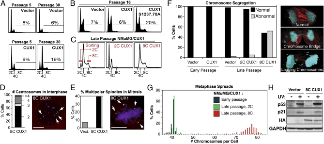
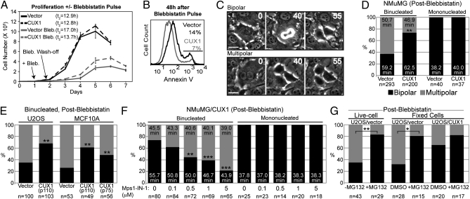
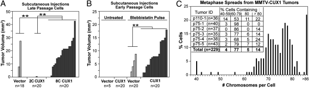
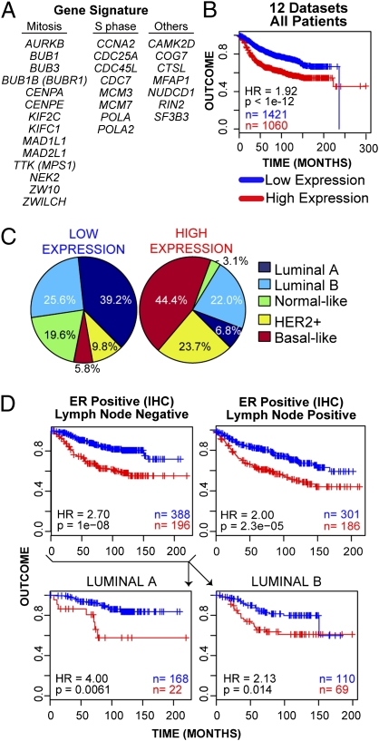
Cell populations able to generate a large repertoire of genetic variants have increased potential to generate tumor cells that survive through the multiple selection steps involved in tumor progression. A mechanism for the generation of aneuploid cancer cells involves passage through a tetraploid stage. Supernumerary centrosomes, however, can lead to multipolar mitosis and cell death. Using tissue culture and transgenic mouse models of breast cancer, we report that Cut homeobox 1 (CUX1) causes chromosomal instability by activating a transcriptional program that prevents multipolar divisions and enables the survival of tetraploid cells that evolve to become genetically unstable and tumorigenic. Transcriptional targets of CUX1 involved in DNA replication and bipolar mitosis defined a gene expression signature that, across 12 breast cancer gene expression datasets, was associated with poor clinical outcome. The signature not only was higher in breast tumor subtypes of worse prognosis, like the basal-like and HER2(+) subtypes, but also identified poor outcome among estrogen receptor-positive/node-negative tumors, a subgroup considered to be at lower risk. The CUX1 signature therefore represents a unique criterion to stratify patients and provides insight into the molecular determinants of poor clinical outcome.


Conflict of interest statement

Conflict of interest statement: L.S., J.L., M.T.H., and A.N. are filing a patent regarding the gene expression signature.

Figures

Fig. 1.
Fig. 1.
Tetraploidy and chromosomal instability in p110 CUX1-expressing NMuMG cells. (A) NMuMG cells stably expressing p110 CUX1 (amino acids 747–1505) were generated by retroviral infection. More than 500 colonies were pooled after selection, which was considered passage 1. Cell cycle distribution was determined by FACS. (B) FACS analysis was performed on independent populations of NMuMG cells expressing either p110 CUX1 or p110S1237,1270A CUX1. (C) Late passage NMuMG/CUX1 cells were FACS-sorted according to DNA content after Hoechst 33342 staining and put back in culture. 2C represent G1 diploid cells and 8C are G2/M tetraploids. (D and E) Number of centrosomes during interphase (D, n = 304) or spindle poles during early mitosis (E, n = 250 for each cell line) was determined by indirect immunofluorescence according to γ-tubulin staining (green), whereas microtubules and DNA were revealed by α-tubulin (red) and DAPI (purple) staining, respectively. (Scale bars, 10 μm.) (F) Chromosome segregation during anaphase was studied by confocal microscopy for late-passage NMuMG/vector (n = 31), early-passage NMuMG/CUX1 (n = 50), and late-passage 2C-sorted (n = 57) 8C-sorted (n = 23) NMuMG/CUX1 cells. Cells were stained for γ-tubulin (green), α-tubulin (red), and DNA (DAPI, blue). Images are a composite of z-sections encompassing entire cells. (G) Chromosome counts were determined on metaphase spreads prepared from early-passage NMuMG/p110 CUX1 or late-passage 2C- or 8C-sorted NMuMG/p110 CUX1 cells (n ≥ 100 each). (H) NMuMG/vector and 8C NMuMG/CUX1 cells were left untreated or exposed to 20 mJ/cm2 UV. Cell lysates were prepared 24 h later and analyzed by SDS/PAGE and Western blotting using the indicated antibodies.
Fig. 2.
Fig. 2.
p110 CUX1 expression enables bipolar mitoses in newly formed tetraploid cells. (A) Proliferation curves of NMuMG/vector (bold) and NMuMG/CUX1 (dashed) cells, with (gray) or without (black) a 12-h blebbistatin pulse treatment. Cells were counted until they ceased proliferation because of contact inhibition. (B) Percentage of apoptotic cells was measured 48 h after blebbistatin wash-off, according to Annexin V staining and FACS analysis. (C and D) Outcome of cell divisions after a blebbistatin wash-off was determined by time-lapse microscopy using frames taken every 5 min. Multipolar divisions displayed a multipolar anaphase giving rise to three or four daughter cells (Movie S1) or a tripolar anaphase followed by cytokinesis failure resulting in two cells, one of which with two nuclei (Movie S2). Neighboring mononucleated cells were used as controls. (Scale bars, 25 μm.) (E) U2OS and MCF10A cells expressing p110 CUX1 were treated with blebbistatin, and the fate of binucleated cells was determined by time-lapse microscopy. (F) NMuMG/CUX1 were pulse-treated with blebbistatin and imaged by time-lapse microscopy with increasing concentrations of MPS1-IN-1. The outcome of cell division and the duration of mitosis were scored for mono- and binucleated cells. (G) U2OS/vector cells were pulse-treated 8 h with blebbistatin. Sixteen hours later, cells were imaged by time-lapse microscopy, during which 10 μM MG132 was added for 90 min and washed away. The fate of binucleated cells entering mitosis during or just before MG132 treatment (Movie S5) was examined. Neighboring binucleated cells dividing without being exposed to MG132 were used as control (Movie S4). Alternatively, cells were fixed 1 h after MG132 treatment, and the metaphase spindle configuration was determined using centrin 3 and α-tubulin staining (Fig. S1 I–K). *P < 0.005; **P < 0.0002; ***P ≤ 1.2 × 10⁻⁵. Black bars, % bipolar division. Gray bars, % multipolar division.
Fig. 3.
Fig. 3.
CUX1-induced tumorigenicity involves chromosomal instability. (A) Late-passage NMuMG/vector cells, 2C-sorted NMuMG/CUX1 cells, or aneuploid 8C-sorted NMuMG/CUX1 cells were injected s.c. in nude mice. Tumor volumes (mm3) were measured 27 d after injection. (B) Early-passage NMuMG cells expressing p110 CUX1 or not were pulse-treated with blebbistatin and injected s.c. into nude mice. (C) Chromosome number of cells from six mammary gland tumors arising in p110 or p75 CUX1 transgenic mice was determined from metaphase spreads. **P < 0.0003 for both tumor sizes and tumor numbers.
Fig. 4.
Fig. 4.
CUX1 transcriptional targets are highly expressed in breast tumor subtypes with poor prognosis and identify a subset of luminal tumors with poor outcome. (A) List of 29 CUX1 transcriptional targets used in expression analysis. Targets include 20 genes validated in Table 1 (but excludes ROD, which is absent from published microarray datasets), plus eight previously characterized DNA replication genes whose expression is strongly stimulated by p110 CUX1 and CTSL1 (12). (B) Kaplan-Meier curve displaying significant segregation of good outcome vs. bad outcome patients classified into “low” and “high” groups on the basis of a 29-gene signature. (C) Breakdown and percentage of patients classified into each molecular subtype for “low” and “high” expression groups. (D) Kaplan-Meier curves comparing the outcome of low- and high-expressing groups for patients reported to be ER+/LN and ER+/LN+. Lower: ER+/LN patients were further classified into luminal A and luminal B subtypes.

References

    1. Rajagopalan H, Lengauer C. Aneuploidy and cancer. Nature. 2004;432:338–341. - PubMed
    1. Fujiwara T, et al. Cytokinesis failure generating tetraploids promotes tumorigenesis in p53-null cells. Nature. 2005;437:1043–1047. - PubMed
    1. Ganem NJ, Godinho SA, Pellman D. A mechanism linking extra centrosomes to chromosomal instability. Nature. 2009;460:278–282. - PMC - PubMed
    1. Quintyne NJ, Reing JE, Hoffelder DR, Gollin SM, Saunders WS. Spindle multipolarity is prevented by centrosomal clustering. Science. 2005;307:127–129. - PubMed
    1. Silkworth WT, Nardi IK, Scholl LM, Cimini D. Multipolar spindle pole coalescence is a major source of kinetochore mis-attachment and chromosome mis-segregation in cancer cells. PLoS ONE. 2009;4:e6564. - PMC - PubMed

Publication types