. 2011 Feb 1;108(5):1914-8.
doi: 10.1073/pnas.1019443108. Epub 2011 Jan 18.

Wild-type LRP6 inhibits, whereas atherosclerosis-linked LRP6R611C increases PDGF-dependent vascular smooth muscle cell proliferation

Affiliations

Wild-type LRP6 inhibits, whereas atherosclerosis-linked LRP6R611C increases PDGF-dependent vascular smooth muscle cell proliferation

Ali R Keramati et al. Proc Natl Acad Sci U S A. .

Abstract

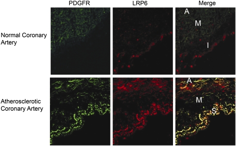
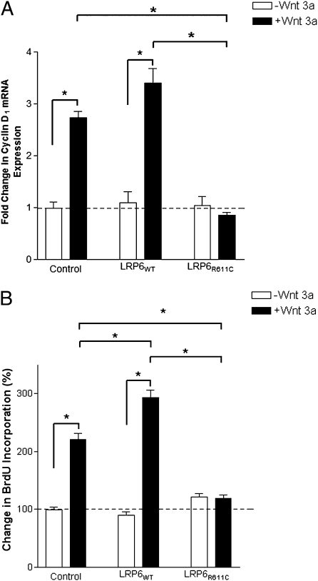
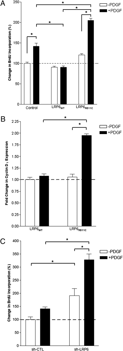
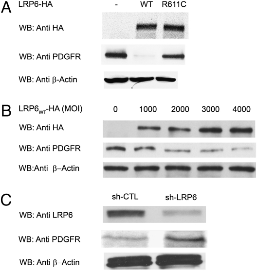
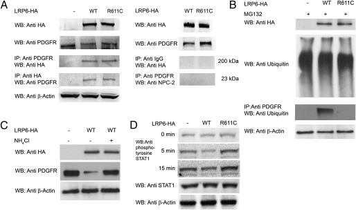
Vascular smooth muscle cell (VSMC) proliferation is an important event in atherosclerosis and other vasculopathies. PDGF signaling is a key mediator of SMC proliferation, but the mechanisms that control its activity remain unclear. We previously identified a mutation in LDL receptor-related protein 6 (LRP6), LRP6(R611C), that causes early atherosclerosis. Examination of human atherosclerotic coronary arteries showed markedly increased expression of LRP6 and colocalization with PDGF receptor β (PDGFR-β). Further investigation showed that wild-type LRP6 inhibits but LRP6(R611C) promotes VSMC proliferation in response to PDGF. We found that wild-type LRP6 forms a complex with PDGFR-β and enhances its lysosomal degradation, functions that are severely impaired in LRP6(R611C). Further, we observed that wild-type and mutant LRP6 regulate cell-cycle activity by triggering differential effects on PDGF-dependent pathways. These findings implicate LRP6 as a critical modulator of PDGF-dependent regulation of cell cycle in smooth muscle and indicate that loss of this function contributes to development of early atherosclerosis in humans.


Conflict of interest statement

The authors declare no conflict of interest.

Figures

Fig. 1.
Fig. 1.
PDGFR-β and LRP6 expression in normal and atherosclerotic coronary arteries. Immunofluorescent staining in a cross-section of a normal coronary artery shows that LRP6 and PDGFR-β are expressed at very low levels in the intima (I) and muscularis (M) layers, respectively (Upper). In the cross-section of the atherosclerotic coronary artery, LRP6 and PDGFR-β are highly expressed and colocalized in the subintima (S) and the muscularis (M) layers (Lower) (n = 5). A, adventicia.
Fig. 2.
Fig. 2.
Effect of LRP6WT and LRP6R611C on cyclin D1 expression and cell proliferation. (A) After 12-h stimulation with Wnt3a, mRNA expression levels of cyclin D1 were about threefold lower in cells expressing LRP6R611C than in uninfected cells (P < 0.0001) or cells expressing LRP6WT (P < 0.001). (B) Wnt3a stimulated the proliferation rate in aortic smooth muscle cells overexpressing LRP6WT and LRP6R611C. Percent BrdU incorporation in cells expressing LRP6R611C after stimulation with Wnt3a remained unchanged and was considerably lower than in cells infected with empty vector (Control) (P < 0.001) or cells expressing LRP6WT (P < 0.001).
Fig. 3.
Fig. 3.
Increased cell-cycle activity in human aortic smooth muscle cells expressing LRP6R611C or LRP6 shRNA. (A) Percent change in BrdU incorporation is considerably higher in cells expressing LRP6R611C in response to PDGF and is considerably lower in cells expressing LRP6WT cells (P < 0.05) than in cells infected with empty vector (P < 0.001). (B) Fold change in cyclin D1 expression levels. After stimulation with PDGF, cyclin D1 expression levels were significantly increased in cells expressing LRP6R611C (P < 0.01) but remained unchanged in cells expressing LRP6WT. (C) Increased proliferation rate in cells infected with LRP6-specific shRNA (sh-LRP6). RNA interference significantly increased the cell proliferation rate both without (P < 0.05) and with PDGF stimulation compared with scrambled shRNA (sh-CTL) (P < 0.001).
Fig. 4.
Fig. 4.
Expression levels of PDGFR-β in human aortic smooth muscle cells expressing LRP6WT and LRP6R611C. (A) LRP6WT significantly reduces PDGFR-β expression levels (P = 0.001). This effect is diminished significantly in the presence of LRP6R611C. (B) Dose effect of LRP6WT on endogenous PDGFR-β expression. There is an inverse relationship between the multiplicity of infection of viral particles expressing LRP6WT-HA2 and PDGFR-β expression. (C) LRP6 knock down by RNA interference (shLRP6). Knocking down LRP6 by RNA interference raised PDGFR-β levels (P = 0.01).
Fig. 5.
Fig. 5.
LRP6WT and LRP6R611C form complexes with PDGFR-β. (A) Proteins from cell lysates were immunoprecipitated with either anti-HA or anti–PDGFR-β antibodies followed by Western blotting with either anti–PDGFR-β or anti-HA antibodies, respectively. LRP6WT and LRP6R611C but not IgG or NPC2 (used as control) coimmunoprecipitated with PDGFR-β. (B) LRP6WT ubiquitinates PDGFR-β. Ubiquitination of PDGFR-β following MG132 treatment in cells overexpressing LRP6R611C or LRP6WT and in cells infected with empty vector was compared. LRP6WT but not LRP6R611C ubiquitinates PDGFR-β. (C) Lysosomal degradation of PDGFR-β. The lysosomal inhibitor NH4Cl rescues reduced expression of PDGFR-β (P = 0.001). (D) LRP6WT reduces and LRP6R611C increases STAT1 tyrosine phosphorylation. After PDGF stimulation, tyrosine phosphorylation of STAT1 is significantly higher in cells expressing LRP6R611C (P < 0.001) and is significantly lower in cells expressing LRP6WT (P < 0.001) than in cells transfected with empty plasmid.

References

    1. Raines EW. PDGF and cardiovascular disease. Cytokine Growth Factor Rev. 2004;15:237–254. - PubMed
    1. Ross R, Glomset JA. The pathogenesis of atherosclerosis (first of two parts) N Engl J Med. 1976;295:369–377. - PubMed
    1. Ross R. The pathogenesis of atherosclerosis: A perspective for the 1990s. Nature. 1993;362:801–809. - PubMed
    1. Ferns GA, et al. Inhibition of neointimal smooth muscle accumulation after angioplasty by an antibody to PDGF. Science. 1991;253:1129–1132. - PubMed
    1. Lewis CD, Olson NE, Raines EW, Reidy MA, Jackson CL. Modulation of smooth muscle proliferation in rat carotid artery by platelet-derived mediators and fibroblast growth factor-2. Platelets. 2001;12:352–358. - PubMed

Publication types

MeSH terms

Substances