. 2011 Feb 1;108(5):1903-7.
doi: 10.1073/pnas.1019267108. Epub 2011 Jan 18.

Membrane lipidome of an epithelial cell line

Affiliations

Membrane lipidome of an epithelial cell line

Julio L Sampaio et al. Proc Natl Acad Sci U S A. .

Abstract

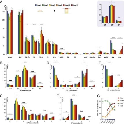
Tissue differentiation is an important process that involves major cellular membrane remodeling. We used Madin-Darby canine kidney cells as a model for epithelium formation and investigated the remodeling of the total cell membrane lipidome during the transition from a nonpolarized morphology to an epithelial morphology and vice versa. To achieve this, we developed a shotgun-based lipidomics workflow that enabled the absolute quantification of mammalian membrane lipidomes with minimal sample processing from low sample amounts. Epithelial morphogenesis was accompanied by a major shift from sphingomyelin to glycosphingolipid, together with an increase in plasmalogen, phosphatidylethanolamine, and cholesterol content, whereas the opposite changes took place during an epithelial-to-mesenchymal transition. Moreover, during polarization, the sphingolipids became longer, more saturated, and more hydroxylated as required to generate an apical membrane domain that serves as a protective barrier for the epithelial sheet.


Conflict of interest statement

The authors declare no conflict of interest.

Figures

Fig. 1.
Fig. 1.
Lipidomics of an EMT. (A) (Lower) EMT was introduced in MDCK B cells transformed with an estrogen-inducible and constitutively active Raf1 mutant and cultured in the presence of estrogen and TGF-β. (Upper) Uninduced cells serve as a control. After 4 d, cells were stained for E-cadherin, actin, and nuclei (DAPI). (B) Lipid class compositions of cells that have undergone an EMT compared with control cells. The content of individual lipid classes was determined by summing up absolute abundances of all identified species and is expressed as mol%. An unpaired t test with Welch's correction was used to estimate the P values: *P < 0.05; **P < 0.01. Error bars correspond to SD (n = 3).
Fig. 2.
Fig. 2.
Time course of polarization. MDCK cells were rendered contact-naive by culturing at low density for 3 d, seeded at low density on filters, and allowed to grow for the indicated time periods. (A) Cells were immunostained for podocalyxin (green), β-catenin (red), and nuclei (blue, DAPI). The x-z views of confocal stacks are shown. (B) Immunoblotting analysis of the respective samples for annexin 13b. Equal loading was confirmed by reprobing for β-actin.
Fig. 3.
Fig. 3.
Lipidomics of polarizing MDCK cells. (A) Lipid class compositions of polarizing MDCK cells; the content of individual lipid classes was determined as described in Fig. 1. (Inset) Cumulative changes within all GPs, SPs, and sterol lipids (ST). Changes in GP chain length, as number of carbon atoms in the sum of fatty acid moieties (B), GP saturation, as number of double bonds in the sum of fatty acid moieties (C), SP chain length, as number of carbon atoms in the sum of long chain base and fatty acid [values for odd numbered chain lengths (n − 1) were summed with values for the even numbered chain lengths (n) depicted] (D), SP saturation, as number of double bonds in the sum of long chain base and fatty acid (E), and SP hydroxylation (F) as calculated from individual quantities of lipid molecular species. (G) Changes in the content of SP species with three hydroxylations within each lipid class; they were not significant for SM but were highly significant for For, GM3, and Sulf (***). P values were estimated by one-way ANOVA (n = 3): *P < 0.05; **P < 0.01; ***P < 0.001. Error bars correspond to SD (n = 3).

References

    1. Thiery JP, Acloque H, Huang RYJ, Nieto MA. Epithelial-mesenchymal transitions in development and disease. Cell. 2009;139:871–890. - PubMed
    1. Nelson WJ. Remodeling epithelial cell organization: Transitions between front-rear and apical-basal polarity. Cold Spring Harb Perspect Biol. 2009;1:a000513. - PMC - PubMed
    1. Leighton J, Estes LW, Mansukhani S, Brada Z. A cell line derived from normal dog kidney (MDCK) exhibiting qualities of papillary adenocarcinoma and of renal tubular epithelium. Cancer. 1970;26:1022–1028. - PubMed
    1. Lever JE. Regulation of dome formation in differentiated epithelial cell cultures. J Supramol Struct. 1979;12:259–272. - PubMed
    1. Bacallao R, et al. The subcellular organization of Madin-Darby canine kidney cells during the formation of a polarized epithelium. J Cell Biol. 1989;109:2817–2832. - PMC - PubMed

Publication types

Substances

LinkOut - more resources