. 2011 Mar;31(6):1121-33.
doi: 10.1128/MCB.01204-10. Epub 2011 Jan 18.

SCF/{beta}-TrCP promotes glycogen synthase kinase 3-dependent degradation of the Nrf2 transcription factor in a Keap1-independent manner

Affiliations

SCF/{beta}-TrCP promotes glycogen synthase kinase 3-dependent degradation of the Nrf2 transcription factor in a Keap1-independent manner

Patricia Rada et al. Mol Cell Biol. 2011 Mar.

Abstract

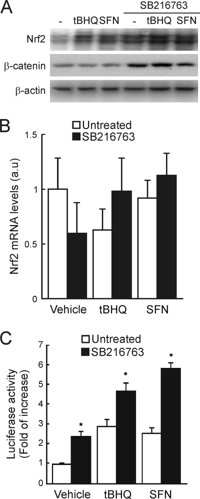
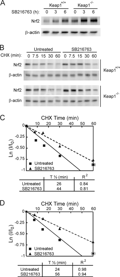
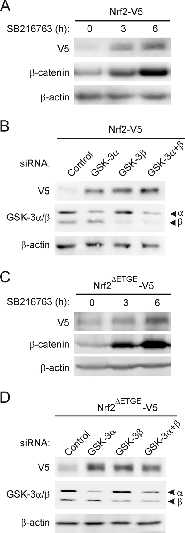
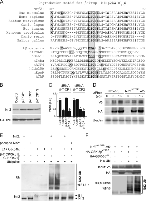
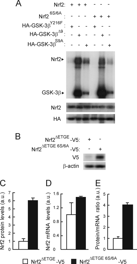
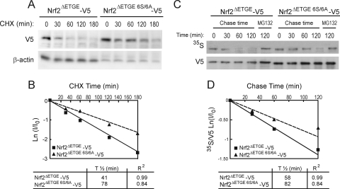
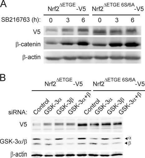
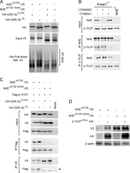
Regulation of transcription factor Nrf2 (NF-E2-related factor 2) involves redox-sensitive proteasomal degradation via the E3 ubiquitin ligase Keap1/Cul3. However, Nrf2 is controlled by other mechanisms that have not yet been elucidated. We now show that glycogen synthase kinase 3 (GSK-3) phosphorylates a group of Ser residues in the Neh6 domain of mouse Nrf2 that overlap with an SCF/β-TrCP destruction motif (DSGIS, residues 334 to 338) and promotes its degradation in a Keap1-independent manner. Nrf2 was stabilized by GSK-3 inhibitors in Keap1-null mouse embryo fibroblasts. Similarly, an Nrf2(ΔETGE) mutant, which cannot be degraded via Keap1, accumulated when GSK-3 activity was blocked. Phosphorylation of a Ser cluster in the Neh6 domain of Nrf2 stimulated its degradation because a mutant Nrf2(ΔETGE 6S/6A) protein, lacking these Ser residues, exhibited a longer half-life than Nrf2(ΔETGE). Moreover, Nrf2(ΔETGE 6S/6A) was insensitive to β-TrCP regulation and exhibited lower levels of ubiquitination than Nrf2(ΔETGE). GSK-3β enhanced ubiquitination of Nrf2(ΔETGE) but not that of Nrf2(ΔETGE 6S/6A). The Nrf2(ΔETGE) protein but not Nrf2(ΔETGE 6S/6A) coimmunoprecipitated with β-TrCP, and this association was enhanced by GSK-3β. Our results show for the first time that Nrf2 is targeted by GSK-3 for SCF/β-TrCP-dependent degradation. We propose a "dual degradation" model to describe the regulation of Nrf2 under different pathophysiological conditions.


Figures

FIG. 1.
FIG. 1.
GSK-3 modulates Nrf2 levels. (A) HEK293T cells were maintained in low-serum medium (0.5% FBS) for 16 h before finally being treated with 15 μM tBHQ, 10 μM SFN, or 20 μM SB216763 for 6 h. Upper blot, Nrf2 immunodetection in cell lysates; middle blot, β-catenin levels as a control for GSK-3 inhibition; lower blot, β-actin levels showing similar protein loads per lane. (B) Quantitative reverse transcriptase PCR (RT-PCR) determination of mRNA of Nrf2 normalized by β-actin from HEK293T cells treated as in panel A (expressed in arbitrary units). Variations are not statistically significant. (C) HEK293T cells were transfected with the 3×ARE-LUC and renilla control vectors, and after transfection the cells were treated with 3 μM tBHQ, 3 μM SFN, or 20 μM SB216763 for 16 h before luciferase activity was measured. Asterisks denote statistically significant differences between the untreated and SB216763-treated groups according to a Student t test.
FIG. 2.
FIG. 2.
GSK-3 inhibition promotes Nrf2 protein accumulation in a Keap1-independent manner. (A) MEFs from Keap1-deficient (Keap1−/−) or wild-type (Keap1+/+) littermates were maintained in low-serum medium for 16 h and then treated with 20 μM SB216763 for the times indicated. Upper blot, total Nrf2 protein levels; lower blot, β-actin levels showing that similar amounts of protein were loaded per lane. (B) MEFs were maintained in low-serum medium for 16 h and then treated with 20 μM SB216763 for 2 h prior to inhibition of protein synthesis with 40 μg/ml cycloheximide (CHX). Whole-cell lysates were prepared at the indicated times after addition of CHX. Upper blots, Nrf2 protein levels in Keap1+/+ and Keap1−/− fibroblasts. Lower blots, β-actin levels showing similar protein loaded per lane from Keap1+/+ and Keap1−/− fibroblasts. (C and D) Both graphs depict the natural logarithm of the relative levels of the Nrf2 protein as a function of CHX chase time in Keap1+/+ (C) or Keap1−/− (D) cells. The protein half-life has been determined in the linear range of the degradation curve.
FIG. 3.
FIG. 3.
Modulation of Nrf2 protein levels by GSK-3 is independent of Keap1. (A and C) HEK293T cells were transfected with either V5-tagged Nrf2, the wild type, or the Keap1-insensitive Nrf2 version (Nrf2ΔETGE-V5), maintained in low-serum medium for 16 h, and then treated with 20 μM SB216763 for the indicated times. Upper blots, Nrf2-V5 (A) or Nrf2ΔETGE-V5 (C) protein levels; middle blots, β-catenin levels in the same cell lysates as a control for GSK-3 inhibition; lower blots, β-actin levels showing similar protein loads per lane. (B and D) HEK293T cells were transfected with either Nrf2-V5 (B) or Nrf2ΔETGE-V5 (D). After 24 h they were further transfected with siRNAs for GSK-3α, GSK-3β, or both or with a control scrambled siRNA as explained in Materials and Methods. Cells were lysed 24 h after siRNA transfection. Upper blots, Nrf2-V5 (B) or Nrf2ΔETGE-V5 (D) protein levels; middle blots, GSK-3α and -β protein levels; lower blots, β-actin levels showing that similar amounts of protein were loaded in each lane.
FIG. 4.
FIG. 4.
Nrf2 is regulated by the E3 ligase β-TrCP complex through a destruction motif within its Neh6 domain. (A) Upper panel of sequences, primary structure of Nrf2 between residues 317 and 359 in the murine protein. The bold/underlined residues correspond to the putative site of phosphorylation by GSK-3, and the boxed ones correspond to the β-TrCP consensus motif. Lower panel of sequences, alignment of different well-known β-TrCP substrates showing the consensus sequence of the degradation motif. (B) Keap1−/− MEFs were transfected with siRNA against β-TrCP1 and/or β-TrCP2 as described in Materials and Methods. Upper blot, Nrf2 total protein levels; lower blot, glyceraldehyde-3-phosphate dehydrogenase (GADPH) levels showing similar protein loads per lane. (C) Quantitative RT-PCR determination of mRNA for β-TrCP1 and β-TrCP2 normalized by β-actin from MEFs transfected as in panel B. Asterisks denote statistically significant differences with P < 0.05. (D) HEK293T cells were cotransfected with the Nrf2-V5 or Nrf2ΔETGE-V5 expression vector and the indicated amounts of the β-TrCP dominant-negative (β-TrCPΔFbox-HA) mutant and then maintained in low-serum medium for 16 h. Whole-cell lysates were immunoblotted against anti-V5 antibody (upper blot) or anti-HA antibody (middle blot) or with anti-β-actin antibody showing similar amounts of protein per sample (lower blot). (E) In vitro ubiquinitation of Nrf2 by the β-TrCP complex. Bacterially expressed His-tagged Nrf2 was submitted to an in vitro kinase assay in the absence (Nrf2) or presence (phospho-Nrf2) of recombinant GSK-3β. Nrf2 and phospho-Nrf2 (20 ng) were incubated at 25°C for 1 h with purified ubiquitin, E1/cdc34b, β-TrCP/Skp1, and Cul1/Rbx1 as indicated. Polyubiquitinated Nrf2 was detected by immunoblotting with antiubiquitin antibody. Upper blot, antiubiquitin. E1-Ub, monoubiquitinated E1. Lower blot, anti-Nrf2 showing similar amounts of substrate loaded per lane. (F) p100 dishes of HEK293T cells were transfected with the indicated plasmids. After transfection (24 h), a whole-cell lysate (input) and an affinity-purified His-tagged fraction (His-pull-down) were blotted with anti-V5 antibody. Upper blot, anti-V5 input; middle blot, ectopically expressed HA-tagged GSK-3β; lower blot, anti-V5 detection in the His pulldown fraction. The bracket indicates the mobility of polyubiquitinated Nrf2-V5 (Nrf2-Ub) forms.
FIG. 5.
FIG. 5.
Mutation of 6S to A in the Neh6 domain of Nrf2 reduces its phosphorylation by GSK-3β. (A) Representative in vitro kinase assay on recombinant Nrf2 or Nrf26S/6A proteins with HA-tagged GSK-3β immunoprecipitated from HEK293T cells transfected with either the inactive HA-GSK-3βY216F mutant or the constitutively active HA-GSK-3βΔ9 or HA-GSK-3βS9A mutant. Upper blot, immunocomplex kinase assay; middle blot, immunodetection of Nrf2 to ensure similar quantity per reaction; lower blot, immunoblot with anti-HA antibody showing similar amount of GSK-3β per assay. (B) Basal levels of Nrf2ΔETGE-V5 or Nrf2ΔETGE 6S/6A-V5 in HEK293T-transfected cells. Upper blot, anti-V5; lower blot, β-actin. (C) Densitometric quantification of representative blots from panel B. (D) Nrf2 mRNA levels analyzed by quantitative RT-PCR from cells transfected as in panel B. (E) Graph depicts the ratio between Nrf2 protein and mRNA levels as shown in panels C and D.
FIG. 6.
FIG. 6.
Mutation of the 6S cluster within the Neh6 domain increases Nrf2 protein stability. (A) HEK293T cells were transfected with Nrf2-ΔETGE-V5 or Nrf2ΔETGE 6S/6A-V5, serum starved for 16 h, and finally subjected to protein synthesis inhibition with 100 μg/ml CHX. Whole-cell lysates were prepared at the indicated times after addition of CHX. Upper blot, Nrf2-V5 protein levels. Lower blot, β-actin levels showing similar protein loads per lane. (B) The graph depicts the natural logarithm of the relative levels of the Nrf2-V5 protein as a function of CHX chase time. The protein half-life was determined using the linear part of the degradation curve. (C) HEK293T cells were transfected with Nrf2-ΔETGE-V5 or Nrf2-ΔETGE 6S/6A-V5, serum starved for 16 h, and then subjected to [35S]methionine/cysteine labeling for 1 h. Then, cells were incubated in high-methionine- and cysteine-containing medium and collected at the indicated times. For the 120-min time point, the cells were treated with MG132 as an internal control. Upper blot, 35S autoradiography; lower blot, anti-V5 antibody. (D) The graph shows the natural logarithm of the relative levels of Nrf2 as a function of 35S chase time. The half-life has been determined in the linear range of the degradation curve.
FIG. 7.
FIG. 7.
Mutation of the 6S cluster within the Neh6 domain renders the Nrf2 protein insensitive to GSK-3. (A) HEK293T cells were transfected with Nrf2-ΔETGE-V5 or Nrf2ΔETGE 6S/6A-V5, maintained in low-serum medium for 16 h, and then incubated with 20 μM SB216763 for the indicated times. Upper blot, anti-V5 immunoblot; middle blot, β-catenin levels as a control of GSK-3 inhibition; lower blot, β-actin levels showing the similar protein loads per lane. (B) HEK293T cells were transfected with either Nrf2ΔETGE-V5 or Nrf2ΔETGE 6S/6A-V5. After 24 h they were further transfected with siRNAs for GSK-3α, GSK-3β, or both or with a control scramble siRNA as described in Materials and Methods. Cells were lysed 24 h after siRNA transfection. Upper blot, anti-V5 antibody; middle blot, GSK-3α and -β protein levels; lower blot, β-actin levels showing similar protein loads per lane.
FIG. 8.
FIG. 8.
The 6S cluster within the Neh6 domain is a target of β-TrCP in a GSK-3-dependent manner. (A) p100 dishes of HEK293T cells were transfected with the indicated plasmids. After transfection (24 h), a whole-cell lysate (input) and an affinity-purified His-tagged fraction (His-Pull-down) were blotted with anti-V5 antibody. Upper panel, anti-V5 input detection; middle panel, ectopically expressed HA-tagged GSK-3β; lower panel, anti-V5 detection in His pulldown fractions. The bracket indicates the mobility of polyubiquitinated Nrf2-V5 (Nrf2-Ub) forms. (B) Keap1−/− or Nrf2−/− MEF cells were serum starved for 6 h before they were treated for a further 2 h with vehicle control, 10 μM LY294002, or 5 μM CT99021. Thereafter, the fibroblasts that had been subjected to different treatments were harvested separately, and each was lysed in 0.45 ml of buffer. A 50-μl portion of the lysate was retained as “input” (labeled on the left), and the remainder was immunoprecipitated with antibodies against either Nrf2 (labeled IP Nrf2) or β-TrCP (labeled IP β-TrCP) using Sepharose-protein G beads. The immunoprecipitated material was analyzed by Western blotting using antibodies against Nrf2 or β-TrCP as indicated to the left of the blots. (C) HEK293T cells were cotransfected with the indicated plasmids or with an empty vector (Mock). One-fifth of whole-protein lysate was used to control for protein expression as shown in the three upper panels. The rest of the protein lysates were immunoprecipitated with anti-Flag or anti-V5 antibodies and immunoblotted as indicated in the four lower panels. The arrow points the specific β-TrCP immunoreactive band. (D) After cotransfection of HEK293T cells with either Nrf2ΔETGE-V5 or Nrf2ΔETGE 6S/6A-V5 and β-TrCPΔFbox-HA, cells were maintained in low-serum medium for 16 h. Upper panel, anti-V5 antibody; middle panel, anti-HA antibody; lower panel, anti-β-actin antibody showing a similar protein load.
FIG. 9.
FIG. 9.
Scheme showing the main participants in the “dual degradation” model. See Discussion for details.

References

    1. Aberle, H., A. Bauer, J. Stappert, A. Kispert, and R. Kemler. 1997. Beta-catenin is a target for the ubiquitin-proteasome pathway. EMBO J. 16:3797-3804. - PMC - PubMed
    1. Apopa, P. L., X. He, and Q. Ma. 2008. Phosphorylation of Nrf2 in the transcription activation domain by casein kinase 2 (CK2) is critical for the nuclear translocation and transcription activation function of Nrf2 in IMR-32 neuroblastoma cells. J. Biochem. Mol. Toxicol. 22:63-76. - PubMed
    1. Calkins, M. J., et al. 2009. The Nrf2/ARE pathway as a potential therapeutic target in neurodegenerative disease. Antioxid. Redox Signal. 11:497-508. - PMC - PubMed
    1. Cuadrado, A., P. Moreno-Murciano, and J. Pedraza-Chaverri. 2009. The transcription factor Nrf2 as a new therapeutic target in Parkinson's disease. Expert Opin. Ther. Targets 13:319-329. - PubMed
    1. Cullinan, S. B., J. D. Gordan, J. Jin, J. W. Harper, and J. A. Diehl. 2004. The Keap1-BTB protein is an adaptor that bridges Nrf2 to a Cul3-based E3 ligase: oxidative stress sensing by a Cul3-Keap1 ligase. Mol. Cell. Biol. 24:8477-8486. - PMC - PubMed

Publication types

MeSH terms