

Nutritional manipulation of primate retinas, V: effects of lutein, zeaxanthin, and n-3 fatty acids on retinal sensitivity to blue-light-induced damage
- PMID: 21245404
- PMCID: PMC3175953
- DOI: 10.1167/iovs.10-5898
Nutritional manipulation of primate retinas, V: effects of lutein, zeaxanthin, and n-3 fatty acids on retinal sensitivity to blue-light-induced damage
Abstract
Purpose: Blue-light photooxidative damage has been implicated in the etiology of age-related macular degeneration (AMD). The macular pigment xanthophylls lutein (L) and zeaxanthin (Z) and n-3 fatty acids may reduce this damage and lower the risk of AMD. This study investigated the effects of the lifelong absence of xanthophylls followed by L or Z supplementation, combined with the effects of n-3 fatty acid deficiency, on acute blue-light photochemical damage.
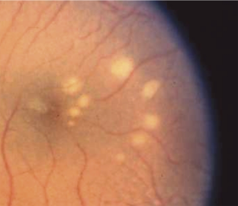
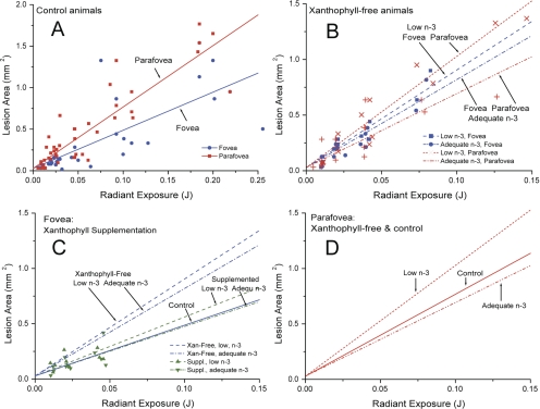
Methods: Subjects included eight rhesus monkeys with no lifelong intake of xanthophylls and no detectable macular pigment. Of these, four had low n-3 fatty acid intake and four had adequate intakes. Control subjects had typical L, Z, and n-3 fatty acid intake. Retinas received 150-μm-diameter exposures of low-power 476-nm laser light at 0.5 mm (∼2°) eccentricity, which is adjacent to the macular pigment peak, and parafoveally at 1.5 mm (∼6°). Exposures of xanthophyll-free animals were repeated after supplementation with pure L or Z for 22 to 28 weeks. Ophthalmoscopically visible lesion areas were plotted as a function of exposure energy, with greater slopes of the regression lines indicating greater sensitivity to damage.
Results: In control animals, the fovea was less sensitive to blue-light-induced damage than the parafovea. Foveal protection was absent in xanthophyll-free animals but was evident after supplementation. In the parafovea, animals low in n-3 fatty acids showed greater sensitivity to damage than animals with adequate levels.
Conclusions: After long-term xanthophyll deficiency, L or Z supplementation protected the fovea from blue light-induced damage, whereas adequate n-3 fatty acid levels reduced the damage in the parafovea.
Figures
References
-
- Ham WT, Mueller HA, Ruffolo JJ, et al. Basic mechanisms underlying the production of photochemical lesions in the mammalian retina. Curr Eye Res. 1984;3:165–174 - PubMed
-
- Ham WT, Allen RG, Feeney-Burns L, Parver LM, Proctor PH, Wolbarscht ML. The involvement of the retinal pigment epithelium (RPE). In: Waxler M, Hitchins VM. eds. Optical Radiation and Visual Health. Boca Raton, FL: CRC Press; 1986:43–67
-
- Dayhaw-Barker P. Ocular photosensitization. Photochem Photobiol. 1987;46:1051–1055 - PubMed
-
- Sparrow JR, Boulton M. RPE lipofuscin and its role in retinal pathobiology. Exp Eye Res. 2005;80:595–606 - PubMed
Publication types
MeSH terms
Substances
Grants and funding
LinkOut - more resources
Full Text Sources
Other Literature Sources
Medical
