. 2011 Feb;121(2):715-25.
doi: 10.1172/JCI43366. Epub 2011 Jan 18.

α-Synuclein propagates from mouse brain to grafted dopaminergic neurons and seeds aggregation in cultured human cells

Affiliations

α-Synuclein propagates from mouse brain to grafted dopaminergic neurons and seeds aggregation in cultured human cells

Christian Hansen et al. J Clin Invest. 2011 Feb.

Abstract

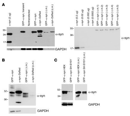
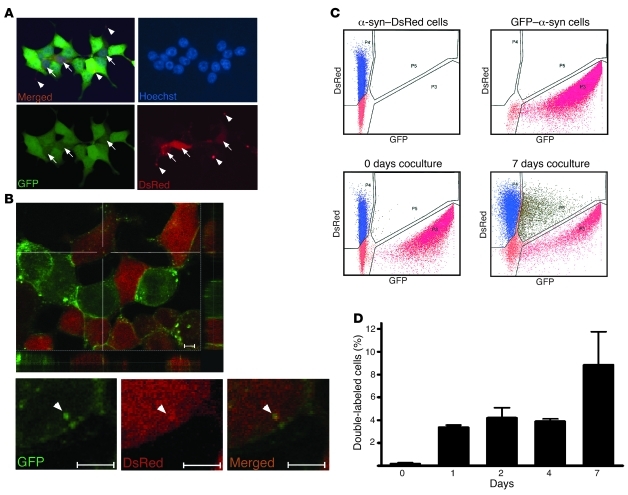
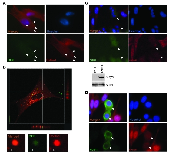
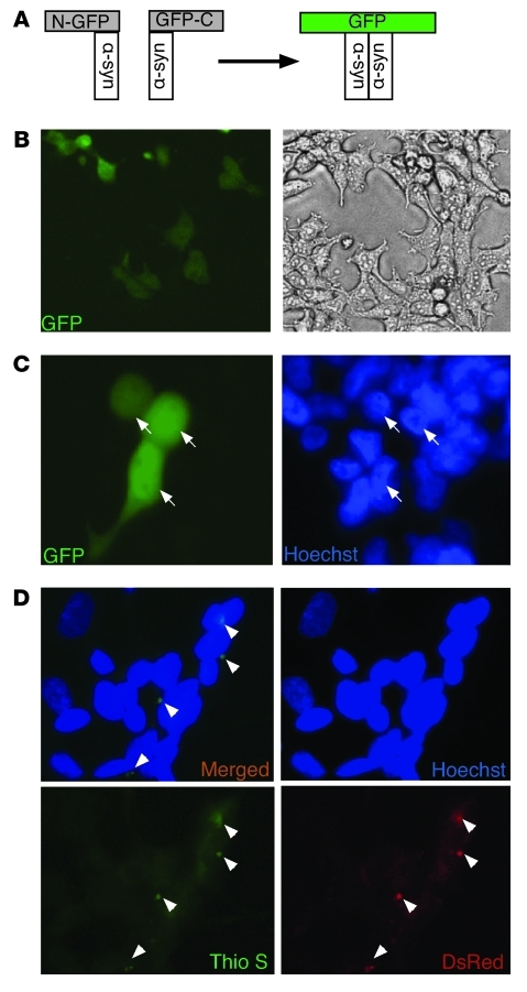
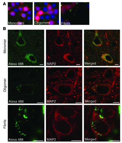
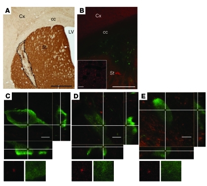
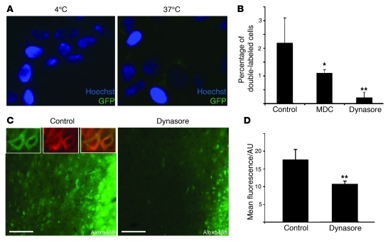
Post-mortem analyses of brains from patients with Parkinson disease who received fetal mesencephalic transplants show that α-synuclein-containing (α-syn-containing) Lewy bodies gradually appear in grafted neurons. Here, we explored whether intercellular transfer of α-syn from host to graft, followed by seeding of α-syn aggregation in recipient neurons, can contribute to this phenomenon. We assessed α-syn cell-to-cell transfer using microscopy, flow cytometry, and high-content screening in several coculture model systems. Coculturing cells engineered to express either GFP- or DsRed-tagged α-syn resulted in a gradual increase in double-labeled cells. Importantly, α-syn-GFP derived from 1 neuroblastoma cell line localized to red fluorescent aggregates in other cells expressing DsRed-α-syn, suggesting a seeding effect of transmitted α-syn. Extracellular α-syn was taken up by cells through endocytosis and interacted with intracellular α-syn. Next, following intracortical injection of recombinant α-syn in rats, we found neuronal uptake was attenuated by coinjection of an endocytosis inhibitor. Finally, we demonstrated in vivo transfer of α-syn between host cells and grafted dopaminergic neurons in mice overexpressing human α-syn. In summary, intercellularly transferred α-syn interacts with cytoplasmic α-syn and can propagate α-syn pathology. These results suggest that α-syn propagation is a key element in the progression of Parkinson disease pathology.


Figures

Figure 1
Figure 1. α-Syn is secreted into the medium of cultured cells.
(A, left) HEK cells not expressing (–) or expressing α-syn fused to GFP or DsRed and 30 μl of conditioned medium from these cells (c.m.). Expression was compared with 2 μg of recombinant α-syn. (A, right) Different concentrations of recombinant α-syn were compared with conditioned medium from 5 different dishes of GFP–α-syn–expressing HEK cells (c.m. 1 to 5). The average concentration of α-syn in the conditioned medium was estimated at 29 ± 3 nM (± SEM). (B) SH-SY5Y cells expressing α-syn fused to GFP or DsRed and 30 μl of conditioned medium from these cells. (C) Comparative Western blots showing expression levels of GFP–α-syn in HEK and SH-SY5Y stable transfectants (left) or in conditioned medium taken from these cells (right). The molecular weights are expressed in kDa on the left side of the blots.
Figure 2
Figure 2. Intercellular propagation of α-syn in HEK cell culture.
(A) Epifluorescence microscopy: representative picture showing double-labeled HEK cells after 5 days of coculture of stable HEK cell lines expressing α-syn fused to either GFP or DsRed. Original magnification, ×40. (B) Confocal microscopy: representative picture showing double-labeled HEK cells after 5 days of coculture of stable HEK cell lines expressing α-syn fused to either GFP or DsRed. Scale bars: 5 μM. Arrows represent double-labeled cells. Arrowheads represent aggregates of transferred α-syn. (C) Representative illustrations of α-syn transfer between cells as evaluated by FACS analysis. Cells expressing α-syn–DsRed or GFP–α-syn were analyzed separately (upper panels) and mixed together just prior to the analysis (negative control, lower left panel) or after 7 days of coculture (lower right panel). (D) Quantification from 20,000 cells in FACS analysis after 0, 1, 2, 4, or 7 days of coculture of α-syn–DsRed– and GFP–α-syn–expressing HEK cells (n = 3). Error bars represent SD.
Figure 3
Figure 3. Intercellular propagation of α-syn in SH-SY5Y cell culture.
(A) Representative picture showing double-labeled SH-SY5Y cells after 14 days of coculture of stable SH-SY5Y cell lines expressing α-syn fused to either GFP or DsRed. Arrows represent transferred aggregates. (B) Confocal microscopy: representative pictures showing double-labeled SH-SY5Y cells after 14 days of coculture of stable SH-SY5Y cell lines expressing α-syn fused to either GFP or DsRed. Scale bars: 2 μM. (C) Upper panel: representative pictures showing double-labeled PC12 cells after 4 days of coculture of human SKMel5 and rat PC12/GFP cells. α-Syn was detected with a monoclonal antibody specific for human α-syn and an Alexa Fluor 568–coupled secondary antibody directed against mouse. Lower panel: comparative Western blot showing the expression level of α-syn in PC12 and SKMel5 cell lysates. Arrows represent double-labeled cells. (D) Representative pictures showing double-labeled N2A cells after 7 days of coculture of human SKMel5 and mouse N2A cells. α-Syn was detected with a monoclonal antibody specific for human α-syn and a Cy3-coupled secondary antibody directed against mouse, and N2A cells were probed with an antibody specific for MAP2 and an Alexa Fluor 488–coupled secondary antibody directed against rabbit. Arrows represent double-labeled cells. Original magnification, ×100 (A); ×40 (C); ×60 (D).
Figure 4
Figure 4. Propagated α-syn interacts with intracellularly expressed α-syn and forms aggregates in recipient cells.
(A) Illustration of the α-syn–based BiFC system. Interaction between 2 α-syn molecules fused to the amino- (N) and carboxyterminal (C) parts of GFP, respectively, leads to reconstitution of GFP fluorescence. (B) HEK cells were transiently transfected with both amino–GFP–α-syn expression construct and carboxy–GFP–α-syn expression construct serving as a positive control. (C) HEK cells were transiently transfected with either amino–GFP–α-syn expression construct or carboxy–GFP–α-syn expression construct and coreplated after 16 hours after media change from OPTIMEM transfection medium to DMEM. GFP fluorescence was monitored after 5 days. Arrows represent GFP-positive cells. (D) Conditioned medium from α-syn–DsRed–expressing HEK cells was incubated with native SH-SY5Y cells for 5 days. Cells were stained with thioflavin S (0.05%) and analyzed for α-syn–DsRed and aggregate formation by fluorescence microscopy. Arrowheads represent DsRed- and Thioflavin S–positive α-syn aggregates. Original magnification, ×10 (B); ×40 (C, D).
Figure 5
Figure 5. In vivo entry of recombinant α-syn proteins into neuronal cells.
(A) 3 hours after addition of 1 μM of Alexa Fluor 488–labeled α-syn proteins in the culture medium, HEK cells expressing α-syn–DsRed have taken up tagged α-syn monomers (left), oligomers (middle), and fibrils (right). Original magnification, ×40. (B) Cortical MAP2-positive neurons internalized Alexa Fluor 488–labeled α-syn monomers (upper panels), oligomers (middle panels), and fibrils (lower panels) after injection into the mouse brain. The left column shows the homogenous distribution of Alexa Fluor 488–labeled (green) monomers throughout the cell body, whereas oligomers and fibrils display a more punctate pattern. The middle column corresponds to the MAP2 staining (red) of recipient neurons and the right column to the merge of left and middle columns. Scale bars: 5 μm.
Figure 6
Figure 6. In vivo transmission of α-syn from mouse brain to a graft of dopaminergic neurons.
(A) Representative coronal section from grafted human α-syn–overexpressing mouse stained with an antibody against TH. The dashed line delineates the transplant of TH-positive neurons in the host striatum. (B) Grafted neurons are identified by TH staining (green) within the human α-syn–positive (red) striatum of the host. The inset shows high magnification of human α-syn–positive accumulations in the host striatum. (CE) Confocal 3D reconstruction of wild-type TH-positive cells (green) in transgenic mice overexpressing human α-syn. The cross points on human α-syn–positive dots (red) present within the transplanted cells. Reconstructed orthogonal projections are presented as viewed in the x-z (bottom) and y-z (right) planes. Cx, cortex; cc, corpus callosum; LV, lateral ventricle; St, striatum. Scale bars: 1,000 μm (A); 500 μm (B); 10 μm (B, inset); 5 μm (CE). Original magnification, ×1915 (CE).
Figure 7
Figure 7. Inhibition of endocytosis decreases α-syn cellular uptake in vitro and in vivo.
(A) Conditioned medium from GFP–α-syn–expressing HEK cells was concentrated 10 times and incubated with native SH-SY5Y cells at 4°C or 37°C. After 6 hours, GFP α-syn uptake was analyzed by fluorescence microscopy. Original magnification, ×40. (B) HEK cells stably transfected with either DsRed- or GFP–tagged α-syn were cocultured for 3 days in the absence (control) or presence of the endocytosis inhibitors monodansylcadaverine (MDC, 1 μM) or dynasore (1 μM), and intercellular transfer was evaluated as percentage of double-labeled cells with epifluorescence microscopy (n = 3). (C) Alexa Fluor 488 signal detected in cortical sections of the same rat injected with Alexa Fluor 488–labeled α-syn monomers on the left side (control) and Alexa Fluor 488–labeled α-syn monomers together with dynasore (80 μM) on the right side (dynasore). The inset in the control picture shows higher magnification of Alexa Fluor 488–positive cells (green), identified all around the injection site of Alexa Fluor 488–labeled α-syn alone. The neuronal nature of these cells is confirmed by colocalization with MAP2 (red). Scale bars: 50 μm. Original magnification, ×60 (top left panels). (D) The mean Alexa Fluor 488 fluorescence per area unit (AU) measured in the injection site, reflecting cellular uptake as a large majority of extracellular Alexa Fluor 488 α-syn monomers, is likely to have been degraded after 6 hours and is decreased by 40% in the dynasore-injected cortex compared with the control side, confirming the inhibition of cellular uptake of Alexa Fluor 488–labeled α-syn monomers in the presence of dynasore. Error bars represent SD. *P < 0.05; **P < 0.01.

References

    1. Spillantini MG, Schmidt ML, Lee VM, Trojanowski JQ, Jakes R, Goedert M. Alpha-synuclein in Lewy bodies. Nature. 1997;388(6645):839–840. doi: 10.1038/42166. - DOI - PubMed
    1. Jakes R, Spillantini MG, Goedert M. Identification of two distinct synucleins from human brain. FEBS Lett. 1994;345(1):27–32. doi: 10.1016/0014-5793(94)00395-5. - DOI - PubMed
    1. Norris EH, Giasson BI, Lee VM. Alpha-synuclein: normal function and role in neurodegenerative diseases. Curr Top Dev Biol. 2004;60:17–54. - PubMed
    1. Burre J, Sharma M, Tsetsenis T, Buchman V, Etherton MR, Sudhof TC. Alpha-synuclein promotes SNARE-complex assembly in vivo and in vitro. Science. 2010;329(5999):1663–1667. doi: 10.1126/science.1195227. - DOI - PMC - PubMed
    1. Kruger R, et al. Ala30Promutation in the gene encoding alpha-synuclein in Parkinson’s disease. Nat Genet. 1998;18(2):106–108. doi: 10.1038/ng0298-106. - DOI - PubMed

Publication types

MeSH terms