Review
. 2011 Jan-Feb;31(1):3-13.
doi: 10.4103/0256-4947.75771.

18F-FDG PET/CT imaging in oncology

Affiliations
Review

18F-FDG PET/CT imaging in oncology

Ahmad Almuhaideb et al. Ann Saudi Med. 2011 Jan-Feb.

Abstract

Accurate diagnosis and staging are essential for the optimal management of cancer patients. Positron emission tomography with 2-deoxy-2-[fluorine-18]fluoro-D-glucose integrated with computed tomography (18F-FDG PET/CT) has emerged as a powerful imaging tool for the detection of various cancers. The combined acquisition of PET and CT has synergistic advantages over PET or CT alone and minimizes their individual limitations. It is a valuable tool for staging and restaging of some tumors and has an important role in the detection of recurrence in asymptomatic patients with rising tumor marker levels and patients with negative or equivocal findings on conventional imaging techniques. It also allows for monitoring response to therapy and permitting timely modification of therapeutic regimens. In about 27% of the patients, the course of management is changed. This review provides guidance for oncologists/radiotherapists and clinical and surgical specialists on the use of 18F-FDG PET/CT in oncology.


Figures

Figure 1
Figure 1
A 73-year-old woman who came for initial staging of non-small cell lung cancer. Maximum-intensity-projection (MIP) image (left panel), CT images (middle panels) and fused images (right panels) of 18F-FDG PET/CT show the primary tumor (arrow head) with mediastinal nodal metastases (black arrow). Incidental right iliac fossa small focal uptake (yellow arrow) is noted, which cross-correlated to a small soft tissue lesion in the cecum and turned out to be a synchronous primary adenocarcinoma.
Figure 2
Figure 2
A 66-year-old woman who came for restaging of breast cancer. MIP image (left panel) and coronal fused images (right panel) of 18F-FDG PET/CT showed extensive hepatic and bony metastases.
Figure 3
Figure 3
A 72-year-old man who had unexplained elevation of CEA during his follow-up after treatment of anorectal cancer. Fused (left panel) and CT (right panel) images of 18F-FDG PET/CT scan showed recurrent avid disease at the residual surgical bed soft tissue density (yellow arrow).
Figure 4
Figure 4
A 56-year-old man who came for initial staging of esophageal cancer. The MIP (left panel) and axial fused (right upper panel) and axial CT (right lower panel) images of 18F-FDG PET/CT showed the primary mid-esophageal tumor with no evidence of FDG-avid distant metastases.
Figure 5
Figure 5
A 68-year-old man who came for initial staging of non-small cell lung cancer. MIP image (left panel) and fused images (right panel) of 18F-FDG PET/CT showed the primary tumor (arrow head) with mediastinal nodal involvement (yellow arrow) and extra-thoracic right adrenal metastasis (black arrow).
Figure 6
Figure 6
A 66-year-old woman diagnosed with Hodgkin lymphoma. The 18F-FDG PET/CT study (left and right upper panels) for initial staging showed nodal involvement above and below the diaphragm. 18F-FDG PET/CT after four cycles of chemotherapy (left and right lower panels) showed complete metabolic resolution of the disease with small non–FDG-avid residual soft tissue (black arrow on the fused image).
Figure 7
Figure 7
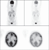
A 62-year-old woman with history of breast cancer. The left column images (MIP and axial PET images) show the normal-intensity images, which could hide metastatic deposits and give a false-negative result due to the physiological high background intensity of the brain. The same images after reducing their intensity on the right column show the metastatic deposits.

References

    1. Yang SK, Cho N, Moon WK. The role of PET/CT for evaluating breast cancer. Korean J Radiol. 2007;8:429–37. - PMC - PubMed
    1. Avril N, Rosé CA, Schelling M, Dose J, Kuhn W, Bense S, et al. Breast imaging with positron emission tomography and fluorine-18 fluorodeoxyglucose: Use and limitations. J Clin Oncol. 2000;18:3495–502. - PubMed
    1. Schirrmeister H, Kühn T, Guhlmann A, Santjohanser C, Hörster T, Nüssle K, et al. Fluorine-18 2-deoxy-2-fluoro-D-glucose PET in the preoperative staging of breast cancer: Comparison with the standard staging procedures. Eur J Nucl Med. 2001;28:351–8. - PubMed
    1. Greco M, Crippa F, Agresti R, Seregni E, Gerali A, Giovanazzi R, et al. Axillary lymph node staging in breast cancer by 2-fluoro-2-deoxy-D-glucose-positron emission tomography: Clinical evaluation and alternative management. J Natl Cancer Inst. 2001;93:630–5. - PubMed
    1. Benard F, Turcotte E. Imaging in breast cancer: Single-photon computed tomography and positron-emission tomography. Breast Cancer Res. 2005;7:153–62. - PMC - PubMed

MeSH terms

Substances