. 2011 Feb 16;30(4):679-91.
doi: 10.1038/emboj.2010.356. Epub 2011 Jan 18.

Membrane surface charge dictates the structure and function of the epithelial Na+/H+ exchanger

Affiliations

Membrane surface charge dictates the structure and function of the epithelial Na+/H+ exchanger

Robert Todd Alexander et al. EMBO J. .

Abstract

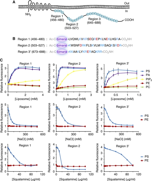
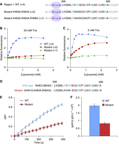
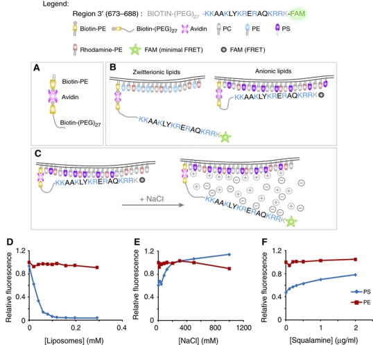
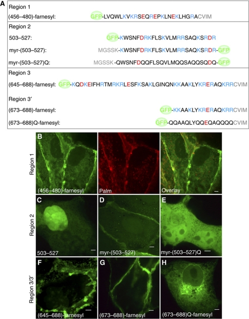
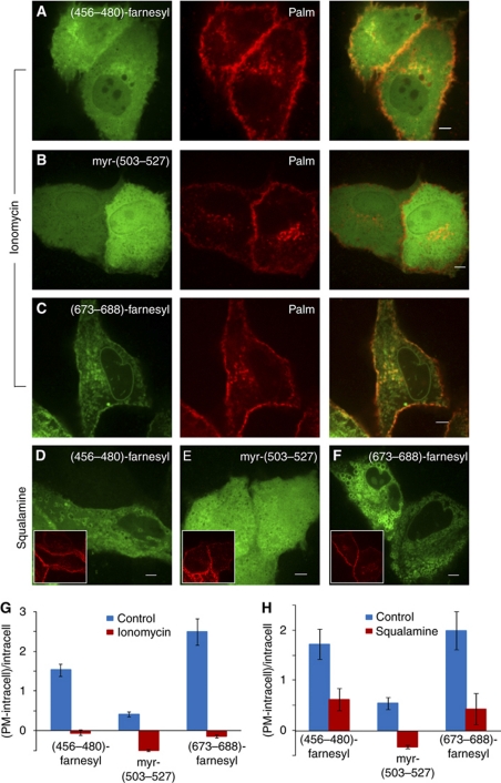
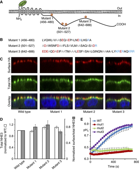
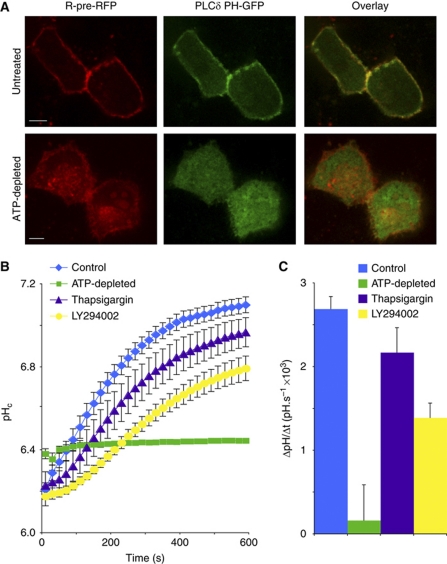
The Na(+)/H(+) exchanger NHE3 plays a central role in intravascular volume and acid-base homeostasis. Ion exchange activity is conferred by its transmembrane domain, while regulation of the rate of transport by a variety of stimuli is dependent on its cytosolic C-terminal region. Liposome- and cell-based assays employing synthetic or recombinant segments of the cytosolic tail demonstrated preferential association with anionic membranes, which was abrogated by perturbations that interfere with electrostatic interactions. Resonance energy transfer measurements indicated that segments of the C-terminal domain approach the bilayer. In intact cells, neutralization of basic residues in the cytosolic tail by mutagenesis or disruption of electrostatic interactions inhibited Na(+)/H(+) exchange activity. An electrostatic switch model is proposed to account for multiple aspects of the regulation of NHE3 activity.


Conflict of interest statement

The authors declare that they have no conflict of interest.

Figures

Figure 1
Figure 1
Cationic regions within the C-terminus of NHE3 bind to liposomes composed of anionic lipids. (A) Representation of NHE3 demonstrating the transmembrane domain and the location of the three cationic regions within the tail of NHE3. (B) Structure of the synthetic cationic peptides labeled with bimane. (C) Graphs of relative bimane fluorescence of a fixed concentration of labeled peptide incubated with increasing concentrations of liposomes (top row), NaCl (middle) or squalamine (bottom). Liposomes contained 20 mol% PS (blue), 20 mol% PA (violet), 2 mol% PIP2 (yellow) and 20 mol% PE (red) with the balance being PC. Liposomes containing only PC are shown in green.
Figure 2
Figure 2
Point mutations depress the binding of region 1 to liposomes and the activity of NHE3. (A) Structure of the bimane-labeled synthetic peptides representing wild-type or mutated versions of region 1. The net charge of the peptides is shown in parentheses. (B, C) Graphs of relative bimane fluorescence of a fixed concentration of labeled peptides incubated with increasing concentrations of liposomes in aqueous medium containing 20 mM Tris–Cl (B) or 5 mM Tris–Cl (C). (D) Sequence of cationic region 1 in wild-type NHE3 (top) and in a triple mutant generated to partially depress the charge of this region. (E) Time course of sodium-induced recovery of pHc in acid-loaded MDCK cells expressing either wild-type or the triple mutant form of NHE3. (F) Rate of pHc recovery in cells expressing the wild-type and mutant forms of NHE3, measured over the first 120 s following addition of sodium. Data are mean values±s.e. of 20 individual determinations of each type.
Figure 3
Figure 3
In vitro model of lipid binding of a membrane-tethered NHE3 domain. (A) Diagram illustrating the means whereby the cationic peptide was tethered to the liposome. (B) Diagram illustrating the predicted location of the peptide relative to the surface of membranes containing zwitterionic or anionic lipids; note the effect of location on probe fluorescence. (C) Putative mechanism whereby increasing ionic strength disrupts the association of the cationic peptide with anionic membranes and the consequences on probe fluorescence. (DF) Graphs of relative fluorescence of a fixed concentration of labeled peptide incubated with varying concentrations of ‘acceptor' liposomes (D), NaCl (E) or squalamine (F). Liposomes contained 20 mol% PS (blue) or 20 mol% PE (red) with 80 mol% PC. Fluorescence in (EF) was normalized to that of PE liposomes.
Figure 4
Figure 4
Expression of cationic domains of NHE3 in mammalian cells. (A) Diagrammatic depiction of the GFP-tagged constructs generated for expression of individual cationic regions. Prenylation and acylation determinants are shown in grey. (B) Representative image showing the distribution of (456–480)-farnesyl (green) in MDCK cells and its colocalization with the plasmalemmal marker Palm construct (red). (CH) Representative images displaying the cellular localization of 503–527 (C), myr-(503–527) (D), myr-(503–527)Q (E), (645–688)-farnesyl (F), (673–688)-farnesyl (G) and (673–688)Q-farnesyl (H) in MDCK cells.
Figure 5
Figure 5
Disruption of the electrostatic interaction between cationic constructs and the plasma membrane results in their release and redistribution. (AC) Representative images of MDCK cells expressing (456–480)-farnesyl (A), myr-(503–527) (B) or (673–688)-farnesyl and the plasmalemmal marker Palm acquired after treatment with ionomycin in the presence of extracellular calcium. (DF) Representative images of MDCK cells expressing (456–480)-farnesyl (D), myr-(503–527) (E) or (673–688)-farnesyl (F) and Palm after treatment with squalamine. Insets show the distribution of Palm. (G, H) Quantification of the association of the indicated constructs with the plasma membrane, calculated as plasmalemmal (PM) fluorescence minus intracellular fluorescence, divided by intracellular fluorescence, before and after treatment with ionomycin (G) or squalamine (H).
Figure 6
Figure 6
Mutation of cationic residues in region 1 or 2 inhibits cation exchange by NHE3. (A, B) Diagrammatic representation of the net charge conferred to regions 1–3 (A) by mutation of cationic residues to alanines, as detailed in (B). (C) Representative XZ reconstructions of surface NHE3 (red), total NHE3 (green) and the nucleus (DAPI/blue) of wild-type and mutant versions of NHE3 expressed in confluent MDCK cells. Size bar=10 μm. (D) Quantification of the total (white bars) and surface (grey bars) expression of three mutants forms of NHE3, relative to the wild type. (E) Sodium-induced recovery of pHc in acid-loaded MDCK cells expressing either wild type or mutants 1–3 of NHE3 (note colour key in the figure inset). Zero second refers to the moment of addition of sodium. Data are mean values±s.e. of at least three experiments of each kind.
Figure 7
Figure 7
Perturbations that disrupt electrostatic interactions with the plasma membrane inhibit NHE3 activity. (A) Subcellular localization of R-pre-RFP (red) and PLCδ-PH-GFP (green) before and after depletion of cellular ATP in MDCK cells. Size bar=5 μm. (B) Sodium-induced recovery of pHc in acid-loaded OK cells (which express endogenous NHE3) that were otherwise untreated (blue), were treated with thapsigargin (purple) or LY294002 (yellow) or subjected to ATP depletion (green). Data are mean values±s.e.m. of at least three determinations of each type. (C) Quantitation of the initial rate of pHc recovery, measured over the first 120 s following addition of sodium in the experiments illustrated in (B).

References

    1. Aharonovitz O, Demaurex N, Woodside M, Grinstein S (1999) ATP dependence is not an intrinsic property of Na+/H+ exchanger NHE1: requirement for an ancillary factor. Am J Physiol 276: C1303–C1311 - PubMed
    1. Aharonovitz O, Zaun HC, Balla T, York JD, Orlowski J, Grinstein S (2000) Intracellular pH regulation by Na+/H+ exchange requires phosphatidylinositol 4,5-bisphosphate. J Cell Biol 150: 213–224 - PMC - PubMed
    1. Akhter S, Cavet ME, Tse CM, Donowitz M (2000) C-terminal domains of Na+/H+ exchanger isoform 3 are involved in the basal and serum-stimulated membrane trafficking of the exchanger. Biochemistry 39: 1990–2000 - PubMed
    1. Akhter S, Nath SK, Tse CM, Williams J, Zasloff M, Donowitz M (1999) Squalamine, a novel cationic steroid, specifically inhibits the brush-border Na+/H+ exchanger isoform NHE3. Am J Physiol 276: C136–C144 - PubMed
    1. Alexander RT, Furuya W, Szaszi K, Orlowski J, Grinstein S (2005) Rho GTPases dictate the mobility of the Na+/H+ exchanger NHE3 in epithelia: role in apical retention and targeting. Proc Natl Acad Sci USA 102: 12253–12258 - PMC - PubMed

Publication types

Substances