. 2011 Jan 18:2:160.
doi: 10.1038/ncomms1157.

Synthetic human cell fate regulation by protein-driven RNA switches

Affiliations
Free PMC article

Synthetic human cell fate regulation by protein-driven RNA switches

Hirohide Saito et al. Nat Commun. .
Free PMC article

Abstract

Understanding how to control cell fate is crucial in biology, medical science and engineering. In this study, we introduce a method that uses an intracellular protein as a trigger for regulating human cell fate. The ON/OFF translational switches, composed of an intracellular protein L7Ae and its binding RNA motif, regulate the expression of a desired target protein and control two distinct apoptosis pathways in target human cells. Combined use of the switches demonstrates that a specific protein can simultaneously repress and activate the translation of two different mRNAs: one protein achieves both up- and downregulation of two different proteins/pathways. A genome-encoded protein fused to L7Ae controlled apoptosis in both directions (death or survival) depending on its cellular expression. The method has potential for curing cellular defects or improving the intracellular production of useful molecules by bypassing or rewiring intrinsic signal networks.


Figures

Figure 1
Figure 1. Schematic diagram for regulation of cell death by protein-driven RNA ON/OFF switches.
(a) Schematic representation of the outcome of this study. A protein encoded in genome drives translational switches and regulates intrinsic apoptosis signal cascade to control cell fate (death or survival). (b) Schematic diagram of the circuit connected to intrinsic apoptosis signal cascades regulated by the switches. Two distinct (mitochondria dependent and independent) apoptosis pathways controlled by L7Ae. L7Ae strictly regulates the production of apoptotic regulatory proteins (that is, Bcl-xL, Bim, FADD) involved in the complex intrinsic apoptosis signal cascades. Several steps of signal-transduction cascades and activation of a set of caspases are required to determine cell death.
Figure 2
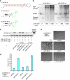
Figure 2. Regulation of the apoptosis pathway using the OFF system.
(a) Schematic illustration of the ON/OFF states of the switch. In cells that do not express L7Ae, translated Bcl-xL protein binds to Bim to prevent apoptosis (ON state). L7Ae represses translation of Bcl-xL, and the apoptosis signal from Bim can proceed (OFF state). (b) Western blotting analysis of Bcl-xL expression in HeLa cells co-transfected with pL7Ae and p(d)Kt-Bcl-xL-I-GFP. Bcl-xL and L7Ae were detected using anti-Bcl-xL and anti-myc antibodies, respectively. (c) Flow cytometric analysis performed 24 h after co-transfection with pBim and the plasmids described in b. The data are presented as the mean±s.d. of triplicate experiments. (d) Phase microscopic images of the cells analysed in c. A scale bar represents 200 μm.
Figure 3
Figure 3. Protein-driven ON system.
(a) Schematic illustration of the ON/OFF states. (b) Sh-GFP targeting the EGFP gene. The sequence in red is complementary to EGFP mRNA (1). Sh-N was designed not to knockdown any gene and encodes a stop codon (in yellow) in every frame (2). We designed Kt-Sh-GFP by incorporating the Kt motif into the loop region of the Sh-GFP (3). dKt-Sh-GFP is defective for the Kt motif in the loop region (4). (c) Determination of the binding affinity of Kt-Sh-GFP (40 nM) and L7Ae (0–640 nM) using the gel shift assay. Kt-Sh-GFP and L7Ae were mixed in transfection buffer, Opti-MEMI (Invitrogen), and separated in the gel shift assay. (d) The specific binding of Kt and L7Ae repressed Dicer cleavage activity for Kt-Sh-GFP. In the presence of L7Ae, Kt-Sh-GFP and L7Ae formed an RNP complex and prevented Dicer cleavage, yet the mutant dKt-Sh-GFP was still cleaved. (e) The relative concentration of EGFP mRNA. (f) The intensity of EGFP fluorescence in HeLa cells stably expressing EGFP (HeLa-GFP). These indicated that L7Ae-specific binding to Kt-Sh-GFP repressed the knock down function of Kt-Sh-GFP and, as a result, inhibited EGFP mRNA degradation and reactivated EGFP expression in HeLa-GFP cells. For e and f, the error bar indicates the standard deviation of three independent samples. Sh-GFP and Sh-N were used for positive and negative controls of EGFP knockdown, respectively.
Figure 4
Figure 4. Using the ON system to control cell fate.
(a) Targeting the Bcl-xL gene with shRNAs containing a loop (1), Kt (2, green) and dKt (3, green). The sequence in red is the complementary strand of a region of Bcl-xL mRNA. The cleavage sites of the Dicer enzyme are indicated by arrowheads. (b) The Dicer cleavage assay in vitro. (c) Western blotting analysis of Bcl-xL using the L7Ae-K-turn ON switch in cells. (d) Flow cytometric analysis of cell death performed 24 h after co-transfection with pAsRed2-L7Ae, pBcl-xL, pBim and the corresponding pshRNA. The data are presented as the mean±s.d. of triplicate experiments. (e) Analysis of the morphology of the cells in d by phase microscopy. A scale bar represents 200 μm.
Figure 5
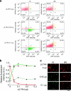
Figure 5. Simultaneous translational repression and activation of two fluorescent proteins by synchronized ON/OFF switches.
(a) Simultaneous translational activation and repression of EGFP and AsRed2 by synchronized ON/OFF switches. The fluorescence intensity of the transfected cells was analysed by flow cytometry. These dot plot data correspond to the fluorescent microscopic images in Figure 5c. (b) Flow cytometric analysis of the simultaneous regulation of EGFP-ON and AsRed2-OFF switches by L7Ae. The efficiencies of translation were normalized to control translational rates in the absence of pL7Ae (AsRed2-OFF) or in the presence of 0.3 μg of pL7Ae (EGFP-ON). The results are presented as the mean±s.d. of triplicate experiments. Green solid line with circles; EGFP relative intensity (Kt), green dashed line with triangles; EGFP relative intensity (dKt), red solid line with circles; AsRed2 relative intensity (Kt), red dashed line with triangles; AsRed2 relative intensity (dKt). (c) Merged fluorescent microscopic images of cells that contained EGFP-ON and AsRed2-OFF switches. A scale bar represents 200 μm.
Figure 6
Figure 6. Simultaneous translational repression and activation of apoptotic proteins by synchronized ON/OFF switches.
(a) Regulation of apoptosis by the synchronized Bcl-xL-ON and Bim-OFF switches. Cells that contain Kt-Bim and Kt-Sh-Bcl-xL were protected from Bim-induced apoptosis by expressing L7Ae (lane 3), whereas cells that contain dKt-Bim and dKt-Sh-Bcl-xL induced apoptosis (lane 6). The cells were analysed by using flow cytometry. Kt-ON/OFF switches are shown in red. The results are presented as the mean±s.d. of triplicate experiments. (b) Cell morphology analysis of Figure 6a by phase microscopy. Scale bars represent 200 μm.
Figure 7
Figure 7. Quantitative regulation of apoptosis by L7Ae-DsRedM protein derived from DNA integrated into genome.
(a) Western blotting analysis of cell lysates 24 h after transfection with pKt-Bcl-xL-I-GFP (or pdKt-Bcl-xL-I-GFP) and pBim in the absence or presence of tetracycline (0–15 ng ml−1) using anti-Bcl-xL antibody. (b) Flow cytometric analysis of the same cells. Cells positive for EGFP and Pacific Blue were considered dead. Blue column, Kt; yellow column, dKt. The results are presented as the mean±s.d. of triplicate experiments.

References

    1. Lu R. et al.. Systems-level dynamic analyses of fate change in murine embryonic stem cells. Nature 462, 358–362 (2009). - PMC - PubMed
    1. Keene J. D. RNA regulons: coordination of post-transcriptional events. Nat. Rev. Genet. 8, 533–543 (2007). - PubMed
    1. Sonenberg N. & Hinnebusch A. G. Regulation of translation initiation in eukaryotes: mechanisms and biological targets. Cell 136, 731–745 (2009). - PMC - PubMed
    1. Gygi S. P., Rochon Y., Franza B. R. & Aebersold R. Correlation between protein and mRNA abundance in yeast. Mol. Cell Biol. 19, 1720–1730 (1999). - PMC - PubMed
    1. Brockmann R., Beyer A., Heinisch J. J. & Wilhelm T. Posttranscriptional expression regulation: what determines translation rates? PLoS Comput. Biol. 3, e57 (2007). - PMC - PubMed