. 2010 Dec 20:4:135-48.
doi: 10.4137/GRSB.S6065.

Molecular Cloning of the Genes Encoding the PR55/Bβ/δ Regulatory Subunits for PP-2A and Analysis of Their Functions in Regulating Development of Goldfish, Carassius auratus

Affiliations

Molecular Cloning of the Genes Encoding the PR55/Bβ/δ Regulatory Subunits for PP-2A and Analysis of Their Functions in Regulating Development of Goldfish, Carassius auratus

Jun-Qiong Zhao et al. Gene Regul Syst Bio. .

Abstract

The protein phosphatase-2A (PP-2A), one of the major phosphatases in eukaryotes, is a heterotrimer, consisting of a scaffold A subunit, a catalytic C subunit and a regulatory B subunit. Previous studies have shown that besides regulating specific PP-2A activity, various B subunits encoded by more than 16 different genes, may have other functions. To explore the possible roles of the regulatory subunits of PP-2A in vertebrate development, we have cloned the PR55/B family regulatory subunits: β and δ, analyzed their tissue specific and developmental expression patterns in Goldfish ( Carassius auratus). Our results revealed that the full-length cDNA for PR55/Bβ consists of 1940 bp with an open reading frame of 1332 nucleotides coding for a deduced protein of 443 amino acids. The full length PR55/Bδ cDNA is 2163 bp containing an open reading frame of 1347 nucleotides encoding a deduced protein of 448 amino acids. The two isoforms of PR55/B display high levels of sequence identity with their counterparts in other species. The PR55/Bβ mRNA and protein are detected in brain and heart. In contrast, the PR55/Bδ is expressed in all 9 tissues examined at both mRNA and protein levels. During development of goldfish, the mRNAs for PR55/Bβ and PR55/Bδ show distinct patterns. At the protein level, PR55/Bδ is expressed at all developmental stages examined, suggesting its important role in regulating goldfish development. Expression of the PR55/Bδ anti-sense RNA leads to significant downregulation of PR55/Bδ proteins and caused severe abnormality in goldfish trunk and eye development. Together, our results suggested that PR55/Bδ plays an important role in governing normal trunk and eye formation during goldfish development.

Keywords: PP-2A; PR55/Bβ/δ; developmental regulation; eye; gene expression; lens; phosphorylation; protein phosphatase.


Figures
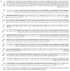
Figure 1
Figure 1
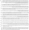
A) The full length cDNA of PR55/Bβ for PP-2A and the deduced protein sequences. The initiation and termination codons are underlined. The predicted amino acid sequence is shown in the one-letter code below the nucleotide sequences. The WD-40 repeats were high-lighted by open box. B) Alignment of the deduced amino acid sequence from goldfish PR55/Bβ of PP-2A with the known PR55/Bβ sequences of PP-2A from human (Homo sapiens, NM_004576), mouse (Mus musculus, NM_088979) and frog (Xenopus laevis, BC130184).
Figure 1
Figure 1
A) The full length cDNA of PR55/Bβ for PP-2A and the deduced protein sequences. The initiation and termination codons are underlined. The predicted amino acid sequence is shown in the one-letter code below the nucleotide sequences. The WD-40 repeats were high-lighted by open box. B) Alignment of the deduced amino acid sequence from goldfish PR55/Bβ of PP-2A with the known PR55/Bβ sequences of PP-2A from human (Homo sapiens, NM_004576), mouse (Mus musculus, NM_088979) and frog (Xenopus laevis, BC130184).
Figure 2
Figure 2
A) The full length cDNA of PR55/Bδ for PP-2A and the deduced protein sequences. The initiation and termination codons are underlined. The predicted amino acid sequence is shown in the one-letter code below the nucleotide sequences. The WD-40 repeats were high-lighted by open box. B) Alignment of the deduced amino acid sequence from goldfish PR55/Bδ of PP-2A with the known PR55/Bδ sequences of PP-2A from Rat (Rattus norvegicus, NM_144746), Mouse (Mus musculus, NM_026391), Chicken (Gallus gallus, NM_001006507), Frog (Xenopus tropicalis, NM_001006696), and Zebrafish (Danio rerio, NM_199776).
Figure 2
Figure 2
A) The full length cDNA of PR55/Bδ for PP-2A and the deduced protein sequences. The initiation and termination codons are underlined. The predicted amino acid sequence is shown in the one-letter code below the nucleotide sequences. The WD-40 repeats were high-lighted by open box. B) Alignment of the deduced amino acid sequence from goldfish PR55/Bδ of PP-2A with the known PR55/Bδ sequences of PP-2A from Rat (Rattus norvegicus, NM_144746), Mouse (Mus musculus, NM_026391), Chicken (Gallus gallus, NM_001006507), Frog (Xenopus tropicalis, NM_001006696), and Zebrafish (Danio rerio, NM_199776).
Figure 3
Figure 3
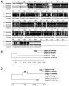
A) Amino acid sequence alignment of the PP2A-PR55/B family members, β/γ/δ in goldfish (The partial amino acid sequence for PR55/Bβ is non-published data from Zhao et al). The completely conserved region among the three isoforms is marked by black shadow. The less conserved region is marked by grey shadow and the non-conserved region is revealed by white background. B) and C) the corresponding phylogenetic trees of the PR55/BβB and PR55/Bδ (C) from four (B) or six (C) vertebrates. The phylogenetic tree for PR55/Bβ (B) was generated through comparative analysis of the coding sequences in human, mouse, frog and the present study using UPGMA calculation and the MEGA3.1 software. The phylogenetic tree for PR55/Bδ (C) was generated using the same strategy and software through comparative analysis of the coding sequences from mouse, rat, chicken, frog, zebrafish and the present study.
Figure 4
Figure 4
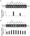
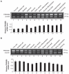
Tissue-specific expression of PR55/Bβ/δ mRNAs of PP-2A in adult goldfish. A) RT-PCR to detect the mRNA level of PR55/Bβ of PP-2A in 9 tissues of adult goldfish as indicated. Up panel: both the PR55/Bβ primers and the β-actin primers were added to the reaction at the beginning of PCR. The 370 bp DNA band for PR55/Bβ mRNA was detected in brain and heart. The 276 bp β-actin DNA band was detected in all tissues. Bottom panel: quantitative results of the PR55/Bβ mRNA levels from three independent experiments. The relative level of expression (fold) was calculated by dividing the total pixel from each PR55/Bβ mRNA band with the total pixel from the corresponding β-actin mRNA band. B) RT-PCR to detect the mRNA level of PR55/Bδ of PP-2A in the same 9 tissues of adult goldfish as indicated in A. Like the 276 bp β-actin DNA band, the 372 bp DNA band for the PR55/Bδ mRNA was also expressed in all the tissues examined. Bottom panel: quantitative results of the PR55/Bδ mRNA expression from three independent experiments. The relative level of expression (fold) was calculated as described above in A.
Figure 5
Figure 5
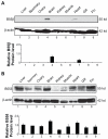
Western blot analysis of the PR55/Bβ and PR55/Bδ proteins in 9 tissues of the adult goldfish indicated. A) Up panel: 100 μg of total proteins extracted from the 9 different tissues of the adult goldfish were subjected to Western blot analysis as described in the Materials and Methods. Bottom panel: quantitative results of PR55/Bβ protein in the above 9 tissues of the adult goldfish from three independent experiments. Note that the highest expression levels of the PR55/Bβ protein was detected in the brain, and to a much less degree in the heart. B) Up panel: 100 μg of total proteins extracted from the 9 different tissues of the adult goldfish were subjected Western blot analysis as described in A. Bottom panel: quantitative results of PR55/Bδ protein in the above 9 tissues of the adult goldfish from three independent experiments. Note that the highest expression levels of the PR55/Bδ protein was detected in the brain and heart, and a reduced level of this protein was detected in liver, spermary, ovary, gin and gill. A much reduced PR55/Bβ/δ protein expression was found in kidney.
Figure 6
Figure 6
Temporal mRNA expression patterns of PR55/Bβ/δ for PP-2A during embryonic development of goldfish. A) Up panel: RT-PCR to detect the mRNA level of PR55/Bβ of PP-2A during 12 different developmental stages. Bottom panel: quantitative results of the PR55/Bβ mRNA expression from three independent experiments. B) Up panel: RT-PCR to detect the mRNA level of PR55/Bδ of PP-2A during the 12 stages. Bottom panel: quantitative results of the PR55/Bδ mRNA expression from three independent experiments. The method is the same as described in Figure 4.
Figure 7
Figure 7
Western blot analysis of the protein for PR55/Bβ/δ in 8 developmental stages as indicated. A) Up panel: 100 micrograms of total proteins extracted from 8 developmental stages of the developing goldfish embryos were subjected to Western blot analysis. Note that no PR55/Bβ protein was detectable at any stage. B) Up panel: 100 micrograms of total proteins extracted from the 8 different stages were subjected to Western blot analysis as described in Figure 5. Bottom panel: quantitative results of PR55/Bδ protein in the 8 developmental stages as determined using the methods described in Figure 5.
Figure 8
Figure 8
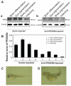
Inhibition of PR55/Bδ protein expression through anti-sense blockage of translation led to severe phenotype in goldfish development. A) The relative expression levels of P55/Bδ in vector-injected (A-a) and anti-sense P55/Bδ expression construct-injected embryos (A-b). B) Quantitation of comparative expression in vector-injected and anti-sense P55/Bδ expression construct-injected embryos. Note that anti-sense blockage of PR55/Bδ translation led to a substantial decrease in the expression level of the PR55/Bδ protein at 4 different stages. The down-regulation of the PR55/Bδ protein caused severe abnormality of organogenesis in the eye and trunk (D) in comparison with the vector-injected embryos (C).

References

    1. Cohen P. The structure and regulation of protein phosphatises. Annu Rev Biochem. 1989;58:453–508. - PubMed
    1. Mumby MC, Walter G. Protein serine/threonine phosphatases: structure, regulation, and functions in cell growth. Physiol Rev. 1993;73:673–99. - PubMed
    1. Hunter T. Protein kinases and phosphatases: the yin and yang of protein phosphorylation and signalling. Cell. 1995;80:225–36. - PubMed
    1. Olsen JV, Blagoev B, Gnad F, et al. Global, in vivo, and site-specific phosphorylation dynamics in signaling networks. Cell. 2006;127:635–48. - PubMed
    1. Moorhead GB, Trinkle-Mulcahy L, Ulke-Lemee A. Emerging roles of nuclear protein phosphatises. Nat Rev Mol Cell Biol. 2007;8:234–44. - PubMed

LinkOut - more resources