. 2011 Jan 5;6(1):e15609.
doi: 10.1371/journal.pone.0015609.

Interaction of Sesbania mosaic virus movement protein with VPg and P10: implication to specificity of genome recognition

Affiliations

Interaction of Sesbania mosaic virus movement protein with VPg and P10: implication to specificity of genome recognition

Soumya Roy Chowdhury et al. PLoS One. .

Abstract

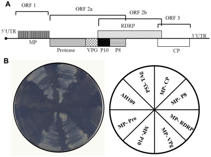
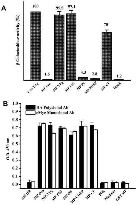
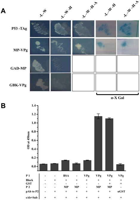
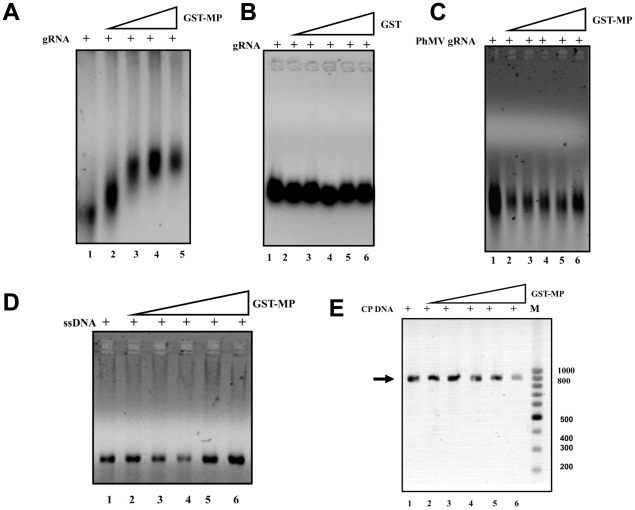
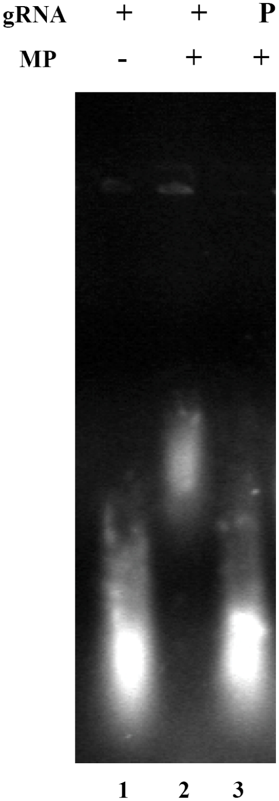
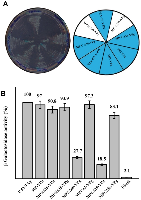
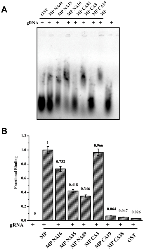
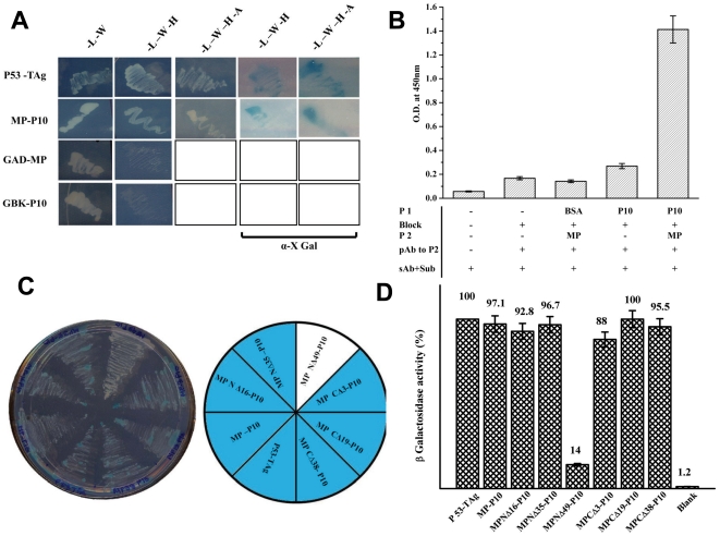
Sesbania mosaic virus (SeMV) is a single strand positive-sense RNA plant virus that belongs to the genus Sobemovirus. The mechanism of cell-to-cell movement in sobemoviruses has not been well studied. With a view to identify the viral encoded ancillary proteins of SeMV that may assist in cell-to-cell movement of the virus, all the proteins encoded by SeMV genome were cloned into yeast Matchmaker system 3 and interaction studies were performed. Two proteins namely, viral protein genome linked (VPg) and a 10-kDa protein (P10) c v gft encoded by OFR 2a, were identified as possible interacting partners in addition to the viral coat protein (CP). Further characterization of these interactions revealed that the movement protein (MP) recognizes cognate RNA through interaction with VPg, which is covalently linked to the 5' end of the RNA. Analysis of the deletion mutants delineated the domains of MP involved in the interaction with VPg and P10. This study implicates for the first time that VPg might play an important role in specific recognition of viral genome by MP in SeMV and shed light on the possible role of P10 in the viral movement.


Conflict of interest statement

Competing Interests: The authors have declared that no competing interests exist.

Figures

Figure 1
Figure 1. Genomic organization of SeMV and Y2H assay for interacting partner of MP.
(A) A schematic representation of the genomic organization of SeMV, representing all the proteins encoded. ORF 1 codes for MP, ORF 2a for poly-protein 2a, ORF 2b for RdRp which is translated by a frame shift mechanism. ORF 3 codes for CP (B) Y2H interaction between MP and all the proteins encoded by SeMV. –Leu –Trp –His plate streaked with AH109 cells transformed with bait and pray plasmids (as shown in the pie diagram). Growth on the plate represents activation of the reporter gene by interaction between the proteins.
Figure 2
Figure 2. Quantitation of Y2H interaction by ß-galactosidase assay and estimation of level of protein expression.
(A) The transformed colonies which showed positive Y2H interaction in the –Leu- Trp- His SD plates were grown in liquid cultures (-Leu –Trp –His medium) and were assayed for β-galactosidase activity to validate and quantify the two-hybrid interactions. For testing the interactions in the pairs that did not grow in -Leu –Trp –His SD plate, the cells grown on -Leu –Trp medium were used. The assay was performed as described in the methods section. The results are presented as the percentage in arbitrary units of ß-galactosidase activity (the values are indicated on the top of each bar) obtained for the interaction between p53 and TAg (100%). The values represent the mean of at least three separate experiments. (B) ELISA to check the expression of the proteins was performed by coating the total protein isolated from AH109 cells transformed with bait and pray plasmid and using c-Myc monoclonal or HA polyclonal antibody as the primary antibody. The bar represents the absorbance measured at 450 nm for each of the protein pairs. The level of expression of all the HA tagged SeMV proteins was comparable to the expression of cMyc tagged MP.
Figure 3
Figure 3. Y2H and ELISA based assays for interaction between MP and VPg.
(A) Y2H interaction between MP and VPg. pGBK T7 (MP and p53 ) and pGAD T7 (VPg and T Ag) clones were transformed in pairs into AH109 strain and plated on to –Leu-Trp SD transformation selection plates and incubated for 96 hrs. Colonies which grew were marked and again replica plated onto various nutritional marker SD plates having different stringency of reporter gene expression as shown in the figure. To determine α- galactosidase activity, colonies were plated onto SD plates having α-X Gal (last two columns). AH109 cells transformed with pairs of pGBK T7-MP and pGAD T7 or pGAD T7-VPg and pGBK T7 clones were also plated on to selection plates to rule out the possibility of auto activation of reporter genes (last two rows). (B) ELISA based interaction study between GST-MP and VPg. VPg (5 µg) (P1) coated ELISA plates were blocked with 10% skimmed milk in 1× PBS (Block) followed by the addition of 5 µg of GST-MP (P2), ELISA was performed with anti MP polyclonal antibody (pAb to P2) and developed using anti rabbit IGg and DMB H2O2 (sAb+Sub). Details of the steps and controls are marked in the figure; BSA and GST were used as the negative controls. The bar represents the absorbance of the samples measured at 450 nm.
Figure 4
Figure 4. Electrophoretic mobility shift assay for binding of nucleic acid substrates to GST-MP.
(A) GST-MP (1, 2, 3 and 4 µg lanes 2–5) was incubated with SeMV genomic RNA (1 µg) in the incubation buffer [200 mM MOPS, 20 mM sodium acetate pH 7] at 4°C for 30 min. The nucleoprotein complex was visualized after electrophoresis on a 0.5% agarose gel, followed by staining with ethidium bromide. Lane 1-RNA alone. (B) Purified GST (1, 2, 3, 4 and 5 µg lanes 2–6) was incubated with SeMV genomic RNA (1 µg) in incubation buffer (as above) at 4°C for 30 min and EMSA was performed as above. Lane 1-RNA alone. (C) Purified GST-MP (1, 2, 3, 4 and 5 µg lanes 2–6) was incubated with PhMV genomic RNA (1 µg) in incubation buffer at 4°C for 30 min and analyzed. Lane 1-RNA alone. (D) GST-MP (1, 2, 3, 4and 5 µg lanes 2–6) was incubated with M13 ssDNA (1 µg) in incubation buffer (as above) at 4°C for 30 min and analysed as before. Lane 1-ssDNA alone. (E) GST-MP (1, 2, 3, 4 and 5 µg lanes 2–6) was incubated with SeMV CP gene PCR product (1 µg) in incubation buffer (as above) at 4°C for 30 min. The nucleoprotein complex was visualized after electrophoresis on a 0.5% agarose gel, followed by staining with ethidium bromide. Lane 1-dsDNA alone.
Figure 5
Figure 5. Role of VPg in the recognition of genomic RNA by MP.
Purified GST-MP (5 μg) was incubated with genomic RNA (1 μg) or pronase treated and trizol purified genomic RNA (1 μg) in incubation buffer [200 mM MOPS, 20 mM sodium acetate pH 7] at 4°C for 30 min. The nucleoprotein complex was visualized after electrophoresis on a 0.5% agarose gel, followed by staining with ethidium bromide. Lane 1- genomic RNA alone, Lane 2- genomic RNA with GST-MP and Lane 3- pronase treated genomic RNA with GST-MP.
Figure 6
Figure 6. Determination of the domains in MP necessary for interaction with VPg by Y2H interaction.
(A) pGBK T7 (MP, MP deletion mutants and p53 ) and pGAD T7 (VPg and T Ag) clones were transformed in pairs into AH109 strain and plated on to –Leu-Trp –His –Ade+α XGal SD high stringency reporter selection plates and incubated for 96 hrs in the dark. Transformants are marked in the pie diagram. (B) Quantification of interaction of MP and MP deletion mutants with VPg by ß-Galactosidase assay. ß-Galactosidase assay of transformed colonies which showed positive Y2H interaction on –Leu –Trp –His –Ade plates was preformed as described in the materials and method section. Data are presented as the percentage of arbitrary units of ß-galactosidase activity (the values are indicated on the top of each bar) with respect to the interaction between p53 and TAg (100%). The values represent the mean of at least three separate experiments.
Figure 7
Figure 7. EMSA of MP and its deletion mutants with viral RNA.
(A) Purified GST, GST MP NΔ49, GST MP NΔ35, GST MP NΔ16, GST MP CΔ38, GST MP CΔ19 and GST MP (5 µg each) were incubated with genomic RNA (1 µg) in incubation buffer [200 mM MOPS, 20 mM sodium acetate pH 7] at 4°C for 30 min. The nucleoprotein complex formed was analysed along with genomic RNA not incubated with MP or its mutants by electrophoresis on a 0.5% agarose gel, followed by staining with ethidium bromide. (B) Fractional binding of RNA was calculated and plotted as a bar diagram for all the proteins using the formula, Fractional binding = Protein bound RNA/ Total RNA.
Figure 8
Figure 8. MP-P10 interaction by Y2H assay.
(A) pGBK T7 (MP and p53 ) and pGAD T7 (P10 and T Ag) clones transformed in pairs into AH109 strain and plated on to –Leu-Trp SD transformation selection plates and incubated for 96 hrs. Colonies which grew were marked and again replica plated on to various nutritional marker SD plates as shown in the figure. To ascertain α- galactosidase activity, colonies were plated onto SD plates having α-X Gal. AH109 cells transformed with pairs of pGBK T7-MP and pGAD T7 or pGAD T7-P10 and pGBK T7 clones were also plated on to selection plates to rule out the possibility of auto activation of reporter genes (last two rows). (B) ELISA based interaction study between GST-MP and P10. P10 (5 µg) (P1) coated ELISA plates were blocked with 10% skimmed milk in 1× PBS (Block) followed by the addition of 5 µg of GST-MP (P2), ELISA was performed with anti MP polyclonal antibody (pAb to P2) and developed using anti rabbit IGg and DMB H2O2 (sAb+Sub). Details of the steps and controls are marked in the figure; BSA was used as the negative control. The bar represents the absorbance of the samples measured at 450nm (C) Determination of the domains in MP necessary for interaction with P10 by Y2H interaction. pGBK T7 (MP, MP deletion mutants and p53 ) and pGAD T7 (P10 and T Ag) clones were transformed in pairs into AH109 strain and plated on to –Leu-Trp –His –Ade +α XGal SD high stringency reporter selection plates and incubated for 96 h in the dark. Transformants are marked in the pie diagram. (D) Quantification of MP and MP deletion mutants – P10 Y2H interaction by ß-galactosidase assay. ß-galactosidase assay of transformed colonies which showed positive Y2H interaction on SD reporter selection plates was performed as described in the methods section. Data are presented as the percentage of arbitrary units of ß-galactosidase activity (the values are indicated on the top of each bar) obtained for the interaction between p53 and TAg (100%). The values represent the mean of at least three separate experiments.

Similar articles

Cited by

References

    1. Carrington JC, Kasschau KD, Mahajan SK, Schaad MC. Cell-to-Cell and Long-Distance Transport of Viruses in Plants. Plant Cell. 1996;8:1669–1681. - PMC - PubMed
    1. Scholthof HB. Plant virus transport: motions of functional equivalence. Trends Plant Sci. 2005;10:376–382. - PubMed
    1. Dolja VV, Haldeman-Cahill R, Montgomery AE, Vandenbosch KA, Carrington JC. Capsid protein determinants involved in cell-to-cell and long distance movement of tobacco etch potyvirus. Virology. 1995;206:1007–1016. - PubMed
    1. Dolja VV, Haldeman R, Robertson NL, Dougherty WG, Carrington JC. Distinct functions of capsid protein in assembly and movement of tobacco etch potyvirus in plants. EMBO J. 1994;13:1482–1491. - PMC - PubMed
    1. Rojas MR, Zerbini FM, Allison RF, Gilbertson RL, Lucas WJ. Capsid protein and helper component-proteinase function as potyvirus cell-to-cell movement proteins. Virology. 1997;237:283–295. - PubMed

Publication types

MeSH terms

Substances