. 2011 Apr;79(4):1215-29.
doi: 10.1002/prot.22957. Epub 2011 Jan 18.

Crystal structure of a bacterial phosphoglucomutase, an enzyme involved in the virulence of multiple human pathogens

Affiliations

Crystal structure of a bacterial phosphoglucomutase, an enzyme involved in the virulence of multiple human pathogens

Ritcha Mehra-Chaudhary et al. Proteins. 2011 Apr.

Abstract

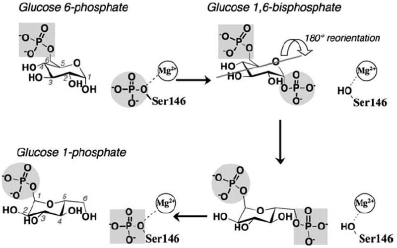
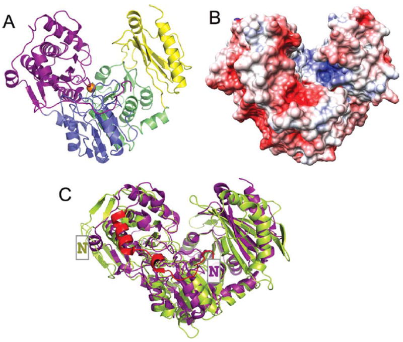
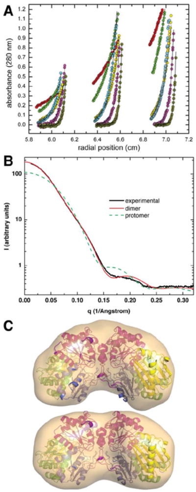
The crystal structure of the enzyme phosphoglucomutase from Salmonella typhimurium (StPGM) is reported at 1.7 A resolution. This is the first high-resolution structural characterization of a bacterial protein from this large enzyme family, which has a central role in metabolism and is also important to bacterial virulence and infectivity. A comparison of the active site of StPGM with that of other phosphoglucomutases reveals conserved residues that are likely involved in catalysis and ligand binding for the entire enzyme family. An alternate crystal form of StPGM and normal mode analysis give insights into conformational changes of the C-terminal domain that occur upon ligand binding. A novel observation from the StPGM structure is an apparent dimer in the asymmetric unit of the crystal, mediated largely through contacts in an N-terminal helix. Analytical ultracentrifugation and small-angle X-ray scattering confirm that StPGM forms a dimer in solution. Multiple sequence alignments and phylogenetic studies show that a distinct subset of bacterial PGMs share the signature dimerization helix, while other bacterial and eukaryotic PGMs are likely monomers. These structural, biochemical, and bioinformatic studies of StPGM provide insights into the large α-D-phosphohexomutase enzyme superfamily to which it belongs, and are also relevant to the design of inhibitors specific to the bacterial PGMs.


Figures

Figure 1
Figure 1
A schematic of the catalytic reaction of StPGM, showing the conversion of G6P to G1P. The bisphosphorylated intermediate undergoes a 180° reorientation in between the two phosphoryl transfer steps of the reaction (gray line indicates axis of rotation). The reaction is highly reversible, thus both G1P and G6P can be considered either as substrates or products.
Figure 2
Figure 2
A: A ribbon diagram of the StPGM monomer colored by domain. Domain 1 (residues 1–205) is shown in purple, domain 2 (residues 206–319) in blue, domain 3 (residues 320–441) in green, and domain 4 (residues 442–546) in yellow. The bound Mg2+ ion in the active site is shown as an orange sphere. B: The electrostatic potential of StPGM shown on a surface rendering. Negative charge is in red, positive in blue. For the purpose of this figure, any charged side chains not included in the crystal structure due to disorder were built in a chemically reasonable position so their charge would contribute to the calculations. Figure made using PMV, MSMS, and Chimera.- C: Ribbon diagram showing a superposition of the monomers of StPGM (purple) and rabbit PGM (green). Chain A from PDB files 3NA5 and 3PMG were used. The N-terminal extension (~20 residues) and dimerization helix of StPGM are shown in red. The NH2-terminus of each protein is indicated by a boxed N.
Figure 3
Figure 3
Biophysical studies of StPGM in solution. A: Sedimentation equilibrium data collected at 6000 (red), 9000 (green), 14,000 (yellow), 12,000 (cyan), 18,000 (magenta), and 25,000 (orange) rpm. The solid lines through the data represent the best fit to a single-species model. For clarity, only a subset of the data points is displayed. B: Experimental and calculated SAXS curves. The thick solid black curve represents the experimental data. The other curves represent SAXS profiles calculated from the dimer (solid red) and monomer (green dashes). C: Shape reconstructions calculated from SAXS data. The surfaces represent the SAXS reconstructions (averaged and filtered volumes) calculated using GASBOR without symmetry constraints (top) and with two-fold symmetry (bottom). The structure of the dimer is shown superimposed onto the SAXS shapes. Superposition calculations were performed using SUPCOMB.
Figure 4
Figure 4
A: Two views of the StPGM dimer as found in the asymmetric unit of crystal form A. Domain 1 of chain A is shown in purple, domain 1 of chain B in cyan. For clarity, the other three domains in each monomer are in white. B: A close-up view of the StPGM dimer interface. Orientation and colors as panel A, top. Side chains of key residues are shown as stick models as follows: the YY motif (red), residues that form hydrogen bonds (green), residues in salt bridges (yellow). C: A close-up view of the N-terminal dimerization helix, viewed down the twofold axis. Proteins are oriented similarly to panel A, bottom. Side chains of interacting residues are colored as in panel B. Only residues in chain A are labeled. D: The proposed C-H⋯π hydrogen bond network involving tyrosines 26 and 27 near the dimer interface of StPGM. Black dashed lines indicate the distance between the hydrogen and the center of mass of the tyrosine rings (red X). Side chains of the involved residues are shown as stick models, with hydrogens (gray) in their calculated positions. Horizontal gray line separates residues in chain A and B of the dimer. Virtually identical interactions (not shown) are found for the same residues in chain B.
Figure 5
Figure 5
A: Close-up view of the active site of StPGM. Domain colors are as Figure 1A. Residues highlighted on Supporting Information Table 3 are shown in stick model and labeled. Dotted line shows the distance in Å between the Cα atoms of the two latch residues (T44 and T511) in form A of StPGM (3NA5). B: A superposition of StPGM (purple) with the closed conformer of parafusin (1KFI) in salmon. Only the phosphate-binding loop of parafusin is shown, highlighting the differences with the same loop in StPGM (see green arrow). In cyan are the bound sulfate molecules found in the 1KFI structure. In green are the side chains of rabbit PGM from regions (i–iii) of the active site. The Mg2+ ion from StPGM is in orange, from rabbit PGM is in green. Also included is the ligand glucose-1,6-bisphosphate (green carbons) from a rabbit PGM structure (1C4G). Despite the presence of the ligand, the rabbit enzyme failed to adopt the closed conformation observed in the sulfate-bound parafusin structure. Nevertheless, the phosphates of the ligand align very closely with the sulfates from the closed parafusin structure, 1KFI, supporting our predicted enzyme–ligand contacts. C: Superposition of the open (purple) and half-closed (cyan) conformers of StPGM, observed in the form A and form B crystal structures respectively. Also superimposed are 17 coordinates sets (gold) showing the conformational fluctuations predicted by a combination of two low frequency normal modes from the elNémo server. The most closed of these 17 conformations closely resembles the sulfate-bound conformer of parafusin, placing the phosphate-binding loop of StPGM in appropriate position to contact bound ligand and close the interdomain latch. D: Residue patches with unusual physicochemical properties in the active sites of (top panel) StPGM (3NA5), and (bottom panel) rabbit PGM (3PMG) highlighting the overall differences between the two proteins. Residue patches (shown as spheres) were calculated by HotPatch, and are colored by rank in decreasing order: red, orange, yellow, green, cyan, and magenta. The locations, residues involved, and number of patches found differ between the two proteins. For both enzymes, the top-ranked (red) patch has a functional confidence value of 1.0, and contains at least three of the key active site residues from Supporting Information Table 3. For StPGM, these are: V371, E390, E391, S392, Y445, R507, and Y518. For rabbit PGM, these are: T18, T356, K359, R502, and R514. Only one identical corresponding residue pair (R507 and R502) is found in this patch for the two proteins. The neural network option in HotPatch that includes a combination of properties (e.g., hydrophobicity, electrostatic potential, etc.) was used for patch identification.

Similar articles

Cited by

References

    1. Paterson GK, Cone DB, Peters SE, Maskell DJ. The enzyme phosphoglucomutase (Pgm) is required by Salmonella enterica serovar Typhimurium for O-antigen production, resistance to antimicrobial peptides and in vivo fitness. Microbiology. 2009;155(Part 10):3403–3410. - PubMed
    1. Ugalde JE, Czibener C, Feldman MF, Ugalde RA. Identification and characterization of the Brucella abortus phosphoglucomutase gene: role of lipopolysaccharide in virulence and intracellular multiplication. Infect Immun. 2000;68:5716–5723. - PMC - PubMed
    1. Bizzini A, Majcherczyk P, Beggah-Moller S, Soldo B, Entenza JM, Gaillard M, Moreillon P, Lazarevic V. Effects of alpha-phosphoglucomutase deficiency on cell wall properties and fitness in Streptococcus gordonii. Microbiology. 2007;153(Part 2):490–498. - PubMed
    1. Grundling A, Schneewind O. Genes required for glycolipid synthesis and lipoteichoic acid anchoring in Staphylococcus aureus. J Bacteriol. 2007;189:2521–2530. - PMC - PubMed
    1. Felek S, Muszynski A, Carlson RW, Tsang TM, Hinnebusch BJ, Krukonis ES. Phosphoglucomutase of Yersinia pestis is required for autoaggregation and polymyxin B resistance. Infect Immun. 2010;78:1163–1175. - PMC - PubMed

Publication types

LinkOut - more resources