. 2011 Feb;240(2):365-71.
doi: 10.1002/dvdy.22541. Epub 2011 Jan 11.

Gpr177/mouse Wntless is essential for Wnt-mediated craniofacial and brain development

Affiliations

Gpr177/mouse Wntless is essential for Wnt-mediated craniofacial and brain development

Jiang Fu et al. Dev Dyn. 2011 Feb.

Abstract

We have previously demonstrated that Gpr177, the mouse orthologue of Drosophila Wls/Evi/Srt, is required for establishment of the anterior-posterior axis. The Gpr177 null phenotype is highly reminiscent to the loss of Wnt3, the earliest abnormality among all Wnt knockouts in mice. The expression of Gpr177 in various cell types and tissues lead us to hypothesize that reciprocal regulation of Wnt and Gpr177 is essential for the Wnt-dependent developmental and pathogenic processes. Here, we create a new mouse strain permitting conditional inactivation of Gpr177. The loss of Gpr177 in the Wnt1-expressing cells causes mid/hindbrain and craniofacial defects which are far more severe than the Wnt1 knockout, but resemble the double knockout of Wnt1 and Wnt3a as well as β-catenin deletion in the Wnt1-expressing cells. Our findings demonstrate the importance of Gpr177 in Wnt1-mediated development of the mouse embryo, suggesting an overlapping function of Wnt family members in the Wnt1-expressing cells.


Figures

Figure 1
Figure 1
Diagram illustrates our targeting strategy and the creation of mice carrying Gpr177Fx and Gpr177Δ allele. (A) In the targeted allele, a loxP site and a pgk-neo cassette flanked by two loxP sites are inserted into intron 2 and intron 3, respectively. Mice carrying the Gpr177 targeted allele were crossed with the EIIa-Cre transgenic mice to generate progeny carrying the Gpr177Fx or Gpr177Δ allele. (B) PCR analysis detects the presence of 5′ (PCR: P1–P2) and 3′ (PCR: P3–P4) loxP sites for genotyping the wild type (+/+), heterozygous (Fx/+) and homozygous (Fx/Fx) mice, and examines the deletion of exon 3 in the Gpr177Δ/+ mice (PCR: P1–P4).
Figure 2
Figure 2
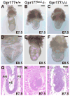
The Gpr177Δ and Gpr177lacZ homozygous embryos exhibit defects in establishment of the body axis. Gross morphological (A–F) and H&E staining (G–I) analyses of the E7.5 (A–C and G–I) and E8.5 (D–F) Gpr177+/+ (A, D, G), Gpr177lacZ−/− (B, E, H) and Gpr177Δ/Δ (C, F, I) embryos reveal that the Gpr177Δ/Δ embryos show deficiencies in patterning of the anterior-posterior axis, identical to those observed in the Gpr177lacZ homozygous embryos. AVE, anterior visceral endoderm; NE, neural ectoderm; PS, primitive streak. Scale bars, 200 μm (A–F); 100 μm (G–I).
Figure 3
Figure 3
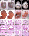
The loss of Gpr177 in the Wnt1-expressing cells causes abnormalities in brain and craniofacial development. Gross morphological (A–H) and H&E staining (I–T) analyses of the E9.5 (A, B), E10.5 (C, D), E13.5 (E, F, I, J) and E16.5 (G, H, K–T) control (genotype: Wnt1-Cre; Gpr177Fx/+ or Gpr177Fx/Fx) and Gpr177Wnt1 (genotype: Wnt1-Cre; Gpr177Fx/Fx) littermates show developmental deformities in the mid/hindbrain and craniofacial structures caused by the deletion of Gpr177 in the Wnt1-expressing cells and their descendants. Arrowhead and arrows indicate the mid/hindbrain boundary and the truncated region, respectively (C, D). Arrows indicate the missing brain structures (I–L). Asterisk indicates cleft palate (M, N). Arrows indicate the tooth, salivary and serous gland defects (O–T). Cb, cerebellum; CP, choroid plexus; Hy, hypothalamus; Inc, incisor; Is, isthmus; My, myelencephalon; Pa, palate; Se, serous gland; SL, sublingual duct; SM, submandibular duct; Tc, tectum; Tg, tegmentum; Th, thalamus. Scale bars, 1 mm (A–D, I, J); 2 mm (E, F, K, L); 4 mm (G, H); 500 μm (M–T).
Figure 4
Figure 4
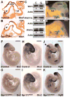
Gpr177 is essential for Wnt-mediated neural development. (A, B) Immunostaining of Wnt1 indicates its expression in the mesencephalon and myelencephalon (arrows) of E9.5 control (genotype: Gpr177Fx/Fx) and Gpr177Wnt1 (genotype: Wnt1-Cre; Gpr177Fx/Fx) embryos. (C) Immunoblot analysis shows the expression levels of Wnt1, Wnt3/3a and Wnt5a in the control (C) and Gpr177Wnt1 (G) mutant embryos at E9.5. The expression level of Actin was analyzed as a loading control. (D, E) Whole mount β-gal staining of E10.5 control (genotype: TOPGAL; Wnt1-Cre; Gpr177Fx/+) and Gpr177Wnt1 (genotype: TOPGAL; Wnt1-Cre; Gpr177Fx/Fx) embryos reveals that deletion of Gpr177 in the Wnt1-expressing cells impairs the expression of TOPGAL transgene in the telencephalon (tel) and mesencephalon (mes). (F–K) Molecular marker analysis of control (genotype: Wnt1-Cre; Gpr177Fx/+ or Gpr177Fx/Fx) and Gpr177Wnt1 (genotype: Wnt1-Cre; Gpr177Fx/Fx) littermates at E9.5 characterizes the effects of the Gpr177 ablation on embryonic brain development using in situ hybridization of En2 (F, G), Otx2 (H, I) and Fgf8 (J, K). Arrowhead indicates isthmic organizer. Scale bars, 200 μm (A, B); 500 μm (D–K).
Figure 5
Figure 5
Development of the neural crest derivatives are impaired by Wnt1-Cre mediated inactivation of Gpr177. (A–J) β-gal staining of E9.5 (A, D), E10.5 (B, E, G–J) and E11.5 (C, F) embryos in whole mounts (A–F) and sections (G–J) examines the neural crest cells which are descendants of the Wnt1-expressing cells in control (genotype: Wnt1-Cre; Gpr177Fx/+; R26RlacZ) and Gpr177Wnt1 (genotype: Wnt1-Cre; Gpr177Fx/Fx; R26RlacZ) littermates. (K, L) In situ hybridization of AP2 labels cranial neural crest cells in the control and Gpr177Wnt1 embryos. (M–P) Neurons of the control and Gpr177Wnt1 embryos were visualized by whole mount immunostaining of neurofilaments in the cranial and trunk regions. Arrowheads indicate the dorsal root ganglions. Cranial nerves: III, oculomotor nerve; V, trigeminal ganglion; VII and VIII, combined ganglion of facial and vestibulocochlear nerves; IX, glossopharyngeal nerve; X, vagus nerve; XII, hypoglossal nerve. (Q–T) Skeletal preparation of the E18.5 control and Gpr177Wnt1 littermates stained with Alizarin Red and Alcian Blue reveals severe abnormalities in the craniofacial skeleton caused by the Gpr177 deletion. Control genotype is Wnt1-Cre; Gpr177Fx/+ or Gpr177Fx/Fx and Gpr177Wnt1 genotype is Wnt1-Cre; Gpr177Fx/Fx. bo, basioccipital bone; bs, basisphenoid; d, mandible; e, exoccipital bone; f, frontal bone; ip, interparietal bone; nc, nasal cartilage; op, olfactory pit; p, parietal bone; ps, presphenoid; px, pre-maxillary bone; s, supraoccipital bone; ty, tympanic ring; x, maxillia; 3v, third ventricle; 4v, fourth ventricle. Scale bars, 500 μm (A–P); 2 mm (Q–T).

References

    1. Brault V, Moore R, Kutsch S, Ishibashi M, Rowitch DH, McMahon AP, Sommer L, Boussadia O, Kemler R. Inactivation of the beta-catenin gene by Wnt1-Cre-mediated deletion results in dramatic brain malformation and failure of craniofacial development. Development. 2001;128:1253–1264. - PubMed
    1. Carpenter AC, Rao S, Wells JM, Campbell K, Lang RA. Generation of mice with a conditional null allele for Wntless. Genesis. 2010;48:554–558. - PMC - PubMed
    1. Chai Y, Jiang X, Ito Y, Bringas P, Jr, Han J, Rowitch DH, Soriano P, McMahon AP, Sucov HM. Fate of the mammalian cranial neural crest during tooth and mandibular morphogenesis. Development. 2000;127:1671–1679. - PubMed
    1. Chiu SY, Asai N, Costantini F, Hsu W. SUMO-Specific Protease 2 Is Essential for Modulating p53-Mdm2 in Development of Trophoblast Stem Cell Niches and Lineages. PLoS Biol. 2008;6:e310. - PMC - PubMed
    1. Clevers H. Wnt/beta-catenin signaling in development and disease. Cell. 2006;127:469–480. - PubMed

Publication types

MeSH terms

LinkOut - more resources