. 2011 May;17(9-10):1375-88.
doi: 10.1089/ten.TEA.2010.0413. Epub 2011 Mar 8.

Mesenchymal stem cell characteristics of human anterior cruciate ligament outgrowth cells

Affiliations

Mesenchymal stem cell characteristics of human anterior cruciate ligament outgrowth cells

Andre F Steinert et al. Tissue Eng Part A. 2011 May.

Abstract

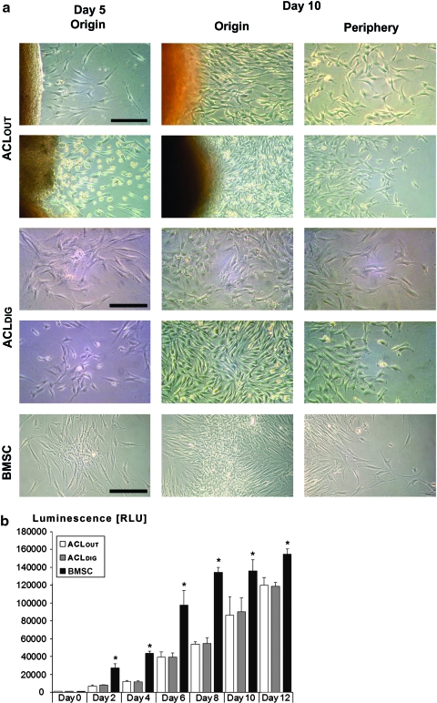
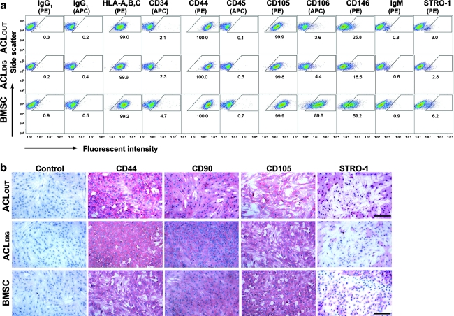
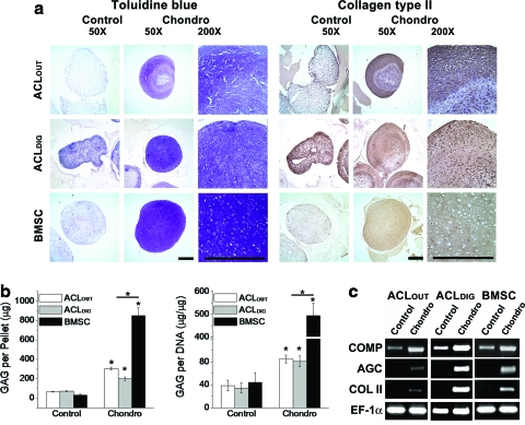
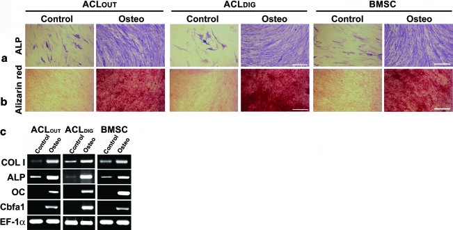
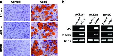
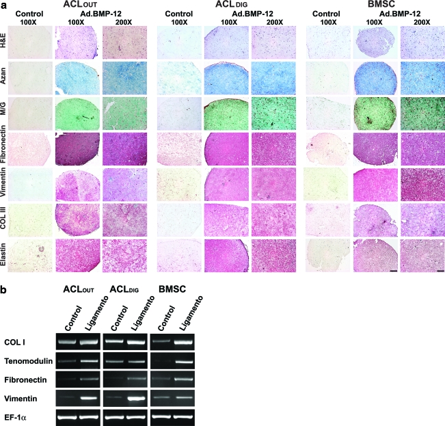
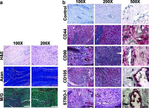
When ruptured, the anterior cruciate ligament (ACL) of the human knee has limited regenerative potential. However, the goal of this report was to show that the cells that migrate out of the human ACL constitute a rich population of progenitor cells and we hypothesize that they display mesenchymal stem cell (MSC) characteristics when compared with adherent cells derived from bone marrow or collagenase digests from ACL. We show that ACL outgrowth cells are adherent, fibroblastic cells with a surface immunophenotype strongly positive for cluster of differentiation (CD)29, CD44, CD49c, CD73, CD90, CD97, CD105, CD146, and CD166, weakly positive for CD106 and CD14, but negative for CD11c, CD31, CD34, CD40, CD45, CD53, CD74, CD133, CD144, and CD163. Staining for STRO-1 was seen by immunohistochemistry but not flow cytometry. Under suitable culture conditions, the ACL outgrowth-derived MSCs differentiated into chondrocytes, osteoblasts, and adipocytes and showed capacity to self-renew in an in vitro assay of ligamentogenesis. MSCs derived from collagenase digests of ACL tissue and human bone marrow were analyzed in parallel and displayed similar, but not identical, properties. In situ staining of the ACL suggests that the MSCs reside both aligned with the collagenous matrix of the ligament and adjacent to small blood vessels. We conclude that the cells that emigrate from damaged ACLs are MSCs and that they have the potential to provide the basis for a superior, biological repair of this ligament.


Figures

FIG. 1.
FIG. 1.
Morphology and cell proliferation of anterior cruciate ligament outgrowth cells (ACLOUT). (a) Phase-contrast photomicrographs of adult human ACLOUT, collagenase-digested ACLDIG cultures, and human bone marrow stromal cells (BMSCs) after 5 and 12 days of culture. Fibroblastic cells migrated from ACL fragments after the first few days of culture (left). Rapidly increasing cell numbers were seen after 12 days, with monolayers reaching confluence after ∼3 weeks (not shown). Two different representative explants are shown for comparison (first and second row). After ACLDIG were placed in culture, where they formed distinct colonies within 5 days. ACLDIG colonies expanded substantially in size by day 12 of culture, and origins and periphery from colonies derived from of two different preparations are shown (third and fourth row). By 5 days after plating bone marrow, BMSCs (bottom row) had formed colonies. These expanded substantially in size by 12 days of culture, and origin and periphery from a representative colony are shown. Scale bar: 100 μm. (b) Comparative cell proliferation rates by means of luminescent measurement of ATP amounts present in all three cell types over time showed increased cell proliferation rates of the BMSCs at all time points compared to the ACLDIG and ACLOUT cultures, which showed no significant differences. Analyses of five different patients are included and n=10 different measurements per cell type, time point, and per patient were performed, with a p-value of 0.5 being considered significant between groups (Asterisks). Color images available online at www.liebertonline.com/tea
FIG. 2.
FIG. 2.
Comparative cell surface phenotying of ACLOUT. (a) Flow cytometric analyses of passage 2 cells revealed that ACLOUT represented nonhematopoietic populations with a very similar surface marker expression profile to that of ACLDIG and BMSCs. In particular, ACLDIG were almost indistinguishable in terms of their surface marker expression compared to the ACLOUT population, with differences in cluster of differentiation (CD)146+ being evident (not significant). However, smaller fractions of both ACL populations stained positively for CD106 and CD146 compared to BMSCs. PE, phycoerythrin; APC, allophycocyanin. Percentage of positive cells for each epitope is given within the corresponding panel. Cell preparations of five individuals for each cell type have been analyzed and representative images are shown. A more comprehensive surface profile comparison is presented in Table 1. (b) Immunocytochemical staining for CD44, CD90, CD105, and STRO-1 in ACLOUT, ACLDIG, and BMSC monolayer cultures revealed strong immunoreactivity (red staining) for the markers CD44, CD90, and CD105. Staining for STRO-1 was also positive, but less widespread as shown in the respective panels, whereas no immunoreactivity was found in the controls. Representative images from cell preparations of 10 donors for each cell type are shown. Scale bar=100 μm. Color images available online at www.liebertonline.com/tea
FIG. 3.
FIG. 3.
Chondrogenic differentiation by ACLOUT versus ACLDIG and BMSC cultures. After 4 weeks in the chondrogenic medium, aggregates derived from ACLOUT, ACLDIG, and BMSCs were analyzed for proteoglycan and type II collagen content as well as for expression of mRNAs associated with chondrogenesis. (a) Paraffin-embedded aggregate sections were stained with Toluidine blue or incubated with anti-human type II collagen antibody (collagen type II). Scale bars=500 μm at both magnifications. Unlike controls, aggregates treated with transforming growth factor beta1 (TGF-β1) (Chondro) from ACLOUT and ACLDIG populations produced an extracellular matrix rich in sulfated proteoglycans and type II collagen similar to the BMSC cultures, although the effect was more homogenous within BMSC aggregates. In proteoglycan- and collagen-rich regions of aggregates, cells were embedded within lacunae-like structures. At least 10 donors for each cell type have been analyzed and representative images are shown. (b) Cells undergoing chondrogenesis produced greater amounts of glycosaminoglycan (GAG) per pellet and per cell compared to controls. This was significantly more pronounced for cells derived from bone marrow. Graphs shown represent results obtained from each of five individual donors, and test have been performed in triplicate for each group and cell type. Asterisks indicate significant differences compared to controls. (c) Aggregate cultures of all three cell types collected at 3 weeks expressed the cartilage-specific genes encoding collagen type II (COL II) and aggrecan core (AGC) protein in response to TGF-β1 treatment, in contrast to controls lacking TGF-β1. Control aggregates expressed cartilage oligomeric matrix protein (COMP) mRNA at low levels, but expression was markedly increased in the presence of TGF-β1. The expression of elongation factor (EF)-1α was included as an internal control for RNA loading. Results are presented using representative patient populations from at least five independent experiments. Color images available online at www.liebertonline.com/tea
FIG. 4.
FIG. 4.
Osteogenic differentiation by ACLOUT versus ACLDIG and BMSC cultures. At least 10 donors for each cell type have been analyzed and representative images are shown. Monolayer cultures were maintained without (Control) and with osteogenic (Osteo) supplements for 3 weeks, and then analyzed by (a, b) cytochemistry and (c) RT-polymerase chain reaction (PCR). (a) Cultures treated with osteogenic supplements contained increased numbers of alkaline phosphatase (ALP)-positive cells (original magnification 200×; bar=100 μm), and (b) produced a mineralized extracellular matrix as shown by intense staining for Alizarin red (original magnification 100×; bar=200 μm). Control cultures contained few ALP+ cells and did not produce a mineralized matrix. Corresponding histomorphometry revealed significant differences of the osteogenic cultures compared to controls for all cell types, but differences between cell types could not be resolved. (c) RT-PCR revealed that ACLOUT as well as ACLDIG and BMSC cultures expressed the osteoblast-related genes osteocalcin (OC), and core binding factor α1 (Cbfa1), in response to osteogenic stimuli, and increased expression of ALP and type I collagen (COL I) compared to controls. Expression of EF-1α was included as an internal control for RNA loading. Color images available online at www.liebertonline.com/tea
FIG. 5.
FIG. 5.
Adipogenic differentiation of ACLOUT versus ACLDIG and BMSC cultures. Monolayer cultures were maintained without (Control) and with adipogenic supplements (Adipo) for 3 weeks, and analyzed for adipogenesis by cytochemistry and RT-PCR. (a) Cultures treated with adipogenic supplements contained cells accumulating large amounts of lipid droplets as shown by intense staining for Oil red-O, in contrast to the respective control cultures (original magnification 200×; scale bar=100 μm), which was significant for all cell types in histomorphometric evaluations (not shown). Differences between cell types were not significant. (b) RT-PCR analysis for expression of lineage-specific genes revealed that ACLOUT, as well as ACLDIG and BMSCs expressed the adipocyte-related lipoprotein lipase (LPL) and proliferator-activator receptor γ2 (PPARγ2) in response to adipogenic medium. Expression of EF-1α was included as a constitutive control for RNA loading. Color images available online at www.liebertonline.com/tea
FIG. 6.
FIG. 6.
Ligamentogenic differentiation of ACLOUT versus ACLDIG and BMSC cultures. As self-renewal is an important stem cell criterion, we employed ACLOUT, ACLDIG, and BMSC populations in an in vitro assay of ligamentogenesis using bone morphogenetic protein 12 (BMP12) encoding adenovirus vectors and three-dimensional culture in collagen hydrogels for differentiation induction compared to controls, which were maintained after marker gene transduction (green fluorescent protein, GFP) in a similar fashion. (a) At 21 days, hematoxylin and eosin (H&E) stainings revealed homogenous cell distribution of fibroblasts for all cell types within the hydrogel constructs (first row), and a strong accumulation of a collagenous matrix after modifications with BMP12 compared to GFP-modified controls where less matrix formation was seen, as evidenced by matrix staining with Azan (second row) and Masson/Goldner (M/G; third row). Immunohistochemical analyses for the ligament matrix proteins fibronectin (fourth row), vimentin (fifth row), collagen type III (Col III; sixth row), and elastin (seventh row) revealed more intense stainings in the Ad.BMP12 transduced hydrogel constructs of all cell types compared to corresponding controls, where no red staining of the matrix was detectable. Differences between cell types (ACLOUT vs. ACLDIG vs. BMSC) could not be detected. Scale bars: 100×bar=200 μm; 200×bar=100 μm. (b) RT-PCR analysis for expression of ligament-specific genes revealed that ACLOUT, as well as ACLDIG and BMSCs, increased expression of the factors COL I, tenomodulin, fibronectin, and vitronectin compared to the respective GFP controls, where only weak expression levels could be detected. Expression of EF-1α was included as a constitutive control for RNA loading. Color images available online at www.liebertonline.com/tea
FIG. 7.
FIG. 7.
Histological and immunohistochemical staining of ACL sections. Pieces of at least n=10 torn and intact ACLs were analyzed and representative images are shown. (a) H&E staining revealed ACL cells within the tissue matrix, as well as the vessel walls. Azan and M/G staining was used to illustrate the density and organization of the collagen fibers that form the tissue matrix within which the ACL cells are embedded. (b) Immunolocalization of antigens CD44, CD90, CD105, and STRO-1 revealed positive cells within the fascicles of the ACL, as well as the endothelium of the vasculature. Examples of such cells are denoted by white (fascicles) and black (endothelium) arrows, respectively, in 500×panels. This indicates that ACL-derived mesenchymal stem cells are likely to originate from both the bulk fascicle tissue as well as the small vessel walls of the ACL. Scale bars: 100×bar=200 μm; 200×bar=100 μm; 500×bar=40 μm. Color images available online at www.liebertonline.com/tea

References

    1. The Burden of Musculoskeletal Diseases in the United States. Rosemont, IL: American Academy of Orthopaedic Surgeons; 2008.
    1. Feagin J.A., Jr. Curl W.W. Isolated tear of the anterior cruciate ligament: 5-year follow-up study. Am J Sports Med. 1976;4:95. - PubMed
    1. Lohmander L.S. Englund P.M. Dahl L.L. Roos E.M. The long-term consequence of anterior cruciate ligament and meniscus injuries: osteoarthritis. Am J Sports Med. 2007;35:1756. - PubMed
    1. Ge Z. Yang F. Goh J.C. Ramakrishna S. Lee E.H. Biomaterials and scaffolds for ligament tissue engineering. J Biomed Mater Res A. 2006;77:639. - PubMed
    1. West R.V. Harner C.D. Graft selection in anterior cruciate ligament reconstruction. J Am Acad Orthop Surg. 2005;13:197. - PubMed

Publication types

MeSH terms

Substances