Review
. 2011 Mar;11(3):254-84.
doi: 10.2174/156800911794519716.

Marizomib, a proteasome inhibitor for all seasons: preclinical profile and a framework for clinical trials

Affiliations
Review

Marizomib, a proteasome inhibitor for all seasons: preclinical profile and a framework for clinical trials

B C Potts et al. Curr Cancer Drug Targets. 2011 Mar.

Abstract

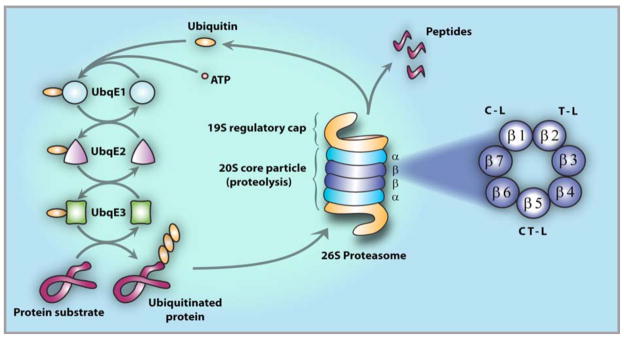
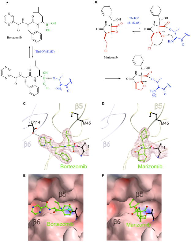
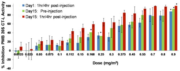
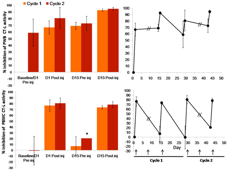
The proteasome has emerged as an important clinically relevant target for the treatment of hematologic malignancies. Since the Food and Drug Administration approved the first-in-class proteasome inhibitor bortezomib (Velcade) for the treatment of relapsed/refractory multiple myeloma (MM) and mantle cell lymphoma, it has become clear that new inhibitors are needed that have a better therapeutic ratio, can overcome inherent and acquired bortezomib resistance and exhibit broader anti-cancer activities. Marizomib (NPI-0052; salinosporamide A) is a structurally and pharmacologically unique β-lactone-γ-lactam proteasome inhibitor that may fulfill these unmet needs. The potent and sustained inhibition of all three proteolytic activities of the proteasome by marizomib has inspired extensive preclinical evaluation in a variety of hematologic and solid tumor models, where it is efficacious as a single agent and in combination with biologics, chemotherapeutics and targeted therapeutic agents. Specifically, marizomib has been evaluated in models for multiple myeloma, mantle cell lymphoma, Waldenstrom's macroglobulinemia, chronic and acute lymphocytic leukemia, as well as glioma, colorectal and pancreatic cancer models, and has exhibited synergistic activities in tumor models in combination with bortezomib, the immunomodulatory agent lenalidomide (Revlimid), and various histone deacetylase inhibitors. These and other studies provided the framework for ongoing clinical trials in patients with MM, lymphomas, leukemias and solid tumors, including those who have failed bortezomib treatment, as well as in patients with diagnoses where other proteasome inhibitors have not demonstrated significant efficacy. This review captures the remarkable translational studies and contributions from many collaborators that have advanced marizomib from seabed to bench to bedside.


Figures

Fig. 1
Fig. 1
The Ubiquitin Protein System (UPS). Protein degradation by the UPS involves two distinct and successive steps: (i) polyubiquitination, i.e., covalent tagging of the target protein with multiple ubiquitin molecules; and (ii) degradation of the tagged protein by the 26S proteasome [3]. Conjugation of ubiquitin to the target protein substrate proceeds via a three-step mechanism, commencing with activation by ubiquitin-activating enzyme, E1, followed by transfer of ubiquitin (via one of several E2 enzymes) from E1 to a member of the ubiquitin-protein ligase family, E3, to which the substrate protein is specifically bound. In successive reactions, a polyubiquitin chain is synthesized by transfer of additional ubiquitin moieties to Lys48 of the previously conjugated molecule. The chain serves as a recognition marker for the 26S proteasome, which degrades the substrates to short peptides by the 20S proteasome and recycles ubiquitin via the action of isopeptidases. The 26S proteasome (center) comprises one or two 19S regulatory caps flanking the proteolytic 20S core particle [22, 23]. The 20S is a cylindrical structure formed by the stacking of two α-rings external to two β-rings, each of which contain seven β subunits, including catalytic subunits β1, β2 and β5 (right, expanded view).
Fig. 2
Fig. 2
20S proteasome inhibition by bortezomib (left panels) and marizomib (right panels). A. Bortezomib forms a non-covalent adduct at the proteasome active site based on the high affinity of boronic acid for the hard oxygen nucleophile Thr1Oγ. The ligand is further stabilized by hydrogen bonding interactions between Thr1NH2 and B-OH, as well as non-covalent P1-P3 residue contacts with the proteasome S1-S3 binding pockets (see panels C, E); the collective binding modality results in slowly reversible proteasome inhibition. B. The β-lactone of marizomib acylates Thr1Oγ, followed by Thr1NH2-catalyzed nucleophilic displacement of chloride by C-3O to give a stable, irreversibly bound adduct. The binding mechanisms for these ligands were established via crystal structures of the yeast 20S proteasome CT-L site (subunit β5) in complex with bortezomib (C, E) and marizomib (D, F). Bortezomib residues P1 and P3 bind to the S1 and S3 pockets, respectively, while boron acts as an electron acceptor for the N-terminal threonine (T1) Thr1Oγ [55]. Marizomib residue P1 binds to the S1 pocket and is covalently bound to T1 via an ester linkage between Thr1Oγ and the carbonyl derived from the β-lactone ring [50]. T1, bortezomib and marizomib are presented as a ball and stick model. Electron density map (mesh) is contoured from 1σ around Thr1 and ligands with 2FO-FC coefficients (C, D). Surface representations of the CT-L active site complex with bortezomib (E) and marizomb (F).
Fig. 3
Fig. 3
Inhibition of the packed whole blood (PWB) CT-L 20S proteasome activity in patient samples increases with dose and is more pronounced after the third administration of marizomib. Marizomib was administered IV on Days 1, 8 and 15 at the doses indicated. Proteasome activity does not restore to baseline levels, as indicated by the inhibition observed on Day 15 before the third administration of marizomib. Results are the average of 3 or more patients per cohort, except where indicated *, the average results of 2 patients is shown.
Fig. 4
Fig. 4
Marizomib pharmacodynamics (percent inhibition of CT-L activity), as monitored in patient PWB and PBMC lysates. Marizomib is administered IV at a dose of 0.55 mg/m2 on Days 1, 8 and 15 of a 28 day cycle. Proteasome activity is assessed before and after the first and third marizomib administration of cycle 1 and 2. Recovery of 20S proteasome CT-L activity between consecutive marizomib administrations is more rapid in PBMCs compared to PWB. Percent inhibition is calculated relative to the Cycle 1 baseline/Day 1 preinjection proteasome activity levels. Results are the average of 3 or more patients, except where indicated *, the average results of 2 patients is shown. Arrow indicates day of marizomib administration.
Fig. 5
Fig. 5
Inhibition of CT-L activity in tumors and various tissues after treatment with marizomib. [52]. MM.1S tumor-bearing mice were injected with three doses of marizomib (0.15 mg/kg, IV, Day 1, Day 4 and Day 8); euthanized at 10 min, 1, 4 and 24 hr after dosing; and PWB, liver, spleen, lung, kidney, brain and tumor were harvested. Protein extracts were prepared and analyzed for CT-L proteasome activity. Results are presented as percent inhibition compared to vehicle control. Data presented are means plus or minus SD (n = 3, p < 0.05).
Fig. 6
Fig. 6
Potential distinct and overlapping mechanisms for synergies between HDAC and proteasome inhibitors.
Fig. 7
Fig. 7
Marizomib treatment reduces tumor burden when added to three conventional colon cancer therapy regimens in a human colon carcinoma (LoVo) xenograft model (n = 6 mice per treatment group, error bars represent the standard error of the mean). CPT-11 (irinotecan); Leuk (leukovorin); Oxa (oxaliplatin); Ava (Avastin; bevacizumab). Adapted with permission from [120].
Fig. 8
Fig. 8
Schematic representation of the involvement of the NF-κB/YY1/Snail/RKIP/DR5 circuit in the regulation of tumor cell resistance to apoptotic stimuli in both chemotherapy and immunotherapy.
Fig. 9
Fig. 9
Marizomib pharmacokinetics. Cmax and AUCtotal dose relationship after single IV administration to cynomolgus monkeys.

References

    1. Adams J. Proteasome Inhibitors in Cancer Therapy. Humana Press; Totowa, NJ: 2004.
    1. Orlowski RZ, Kuhn DJ. Proteasome inhibitors in cancer therapy: lessons from the first decade. Clin Cancer Res. 2008;14:1649–1657. - PubMed
    1. Hershko A, Ciechanover A. The ubiquitin system. Annu Rev Biochem. 1998;67:425–479. - PubMed
    1. Palombella VJ, Rando OJ, Goldberg AL, Maniatis T. The ubiquitin-proteasome pathway is required for processing the NF-kappa B1 precursor protein and the activation of NF-kappa B. Cell. 1994;78:773–785. - PubMed
    1. Chauhan D, Uchiyama H, Akbarali Y, Urashima M, Yamamoto K, Libermann TA, Anderson KC. Multiple myeloma cell adhesion-induced interleukin-6 expression in bone marrow stromal cells involves activation of NF-kappa B. Blood. 1996;87:1104–1112. - PubMed

Publication types

MeSH terms