. 2011 Jan 19;3(1):2.
doi: 10.1186/1758-2555-3-2.

Stretching positions for the coracohumeral ligament: Strain measurement during passive motion using fresh/frozen cadaver shoulders

Affiliations

Stretching positions for the coracohumeral ligament: Strain measurement during passive motion using fresh/frozen cadaver shoulders

Tomoki Izumi et al. Sports Med Arthrosc Rehabil Ther Technol. .

Abstract

Background: Contracture of the coracohumeral ligament is reported to restrict external rotation of the shoulder with arm at the side and restrict posterior-inferior shift of the humeral head. The contracture is supposed to restrict range of motion of the glenohumeral joint.

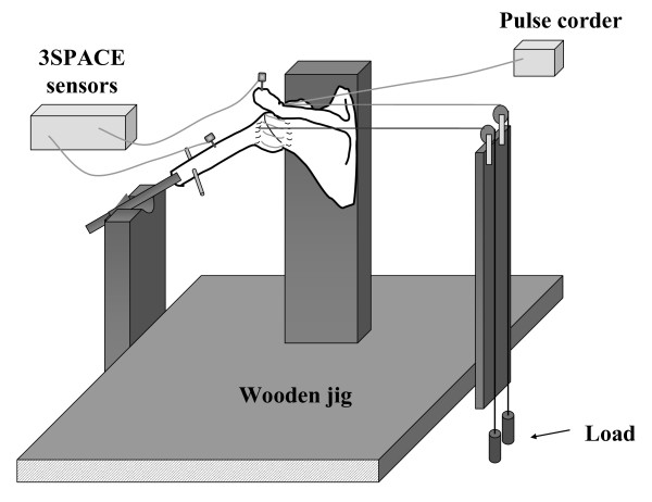
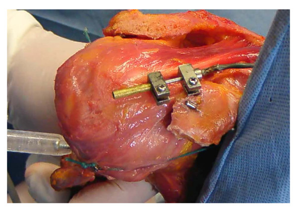
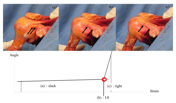
Methods: To obtain stretching position of the coracohumeral ligament, strain on the ligament was measured at the superficial fibers of the ligament using 9 fresh/frozen cadaver shoulders. By sequential measurement using a strain gauge, the ligament strain was measured from reference length (L0). Shoulder positions were determined using a 3 Space Tracker System. Through a combination of previously reported coracohumeral stretching positions and those observed in preliminary measurement, ligament strain were measured by passive external rotation from 10° internal rotation, by adding each 10° external rotation, to maximal external rotation.

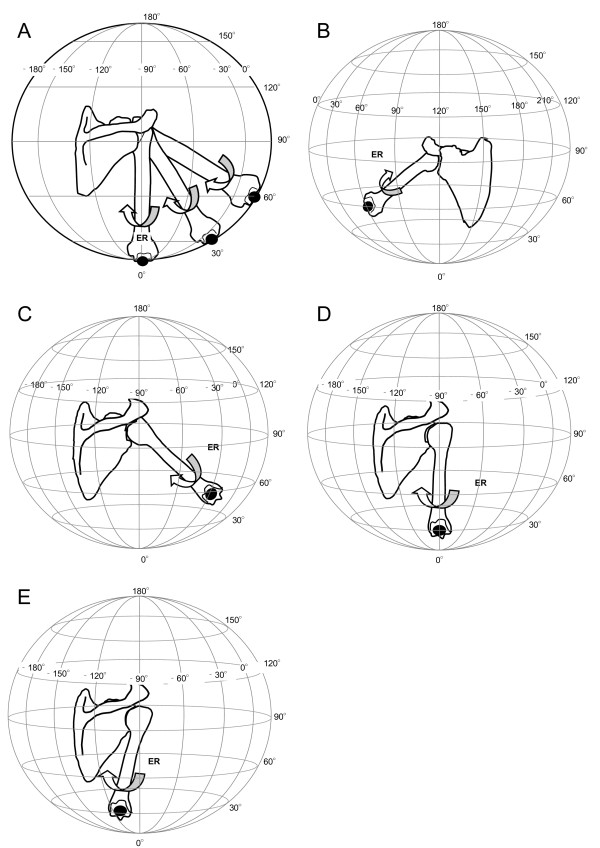
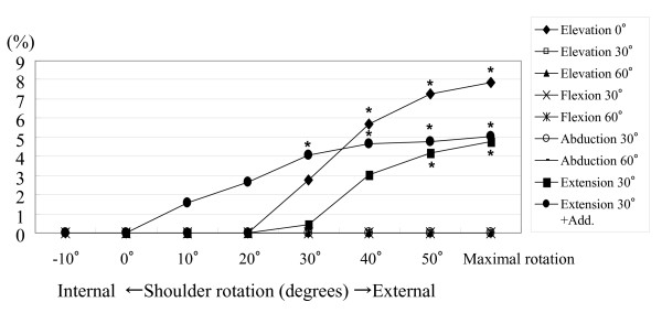
Results: Stretching positions in which significantly larger strain were obtained compared to the L0 values were 0° elevation in scapula plane with 40°, 50° and maximum external rotation (5.68%, 7.2%, 7.87%), 30° extension with 50°, maximum external rotation (4.20%, 4.79%), and 30° extension + adduction with 30°, 40°, 50° and maximum external rotation (4.09%, 4.67%, 4.78%, 5.05%)(P < 0.05). No positive strain on the coracohumeral ligament was observed for the previously reported stretching positions; ie, 90° abduction with external rotation or flexion with external rotation.

Conclusions: Significant strain of the coracohumeral ligament will be achieved by passive external rotation at lower shoulder elevations, extension, and extension with adduction.


Figures

Figure 1
Figure 1
Experimental set-up. A displacement sensor was attached to the superficial fibers of the coracohumeral ligament. A six-degree of freedom electromagnetic tracking device was used to monitor the glenohumeral rotational angles.
Figure 2
Figure 2
A photograph of the displacement sensor. The sensor was attached to the center of the superficial fibers of the coracohumeral ligament parallel to the ligament fibers.
Figure 3
Figure 3
Measurement positions for the glenohumeral joint in vitro. External rotation at 0°, 30° and 60° elevation in the scapula plane (Figure 3-A), external rotation at 30° and 60° flexion (Figure 3-B), external rotation at 30° and 60° abduction (Figure 3-C), external rotation at 30° extension (Figure 3-D), external rotation and adduction at 30° extension (Figure 3-E). In the globe system, the scapula plane was consistent with 0 degree of longitude. Then, the latitude of the globe system indicates elevation angle of the glenohumeral joint. The longitude of globe system indicates horizontal adduction or abduction angle of the glenohumeral joint. Arrows indicated direction of motion of the glenohumeral joint as external rotation. ER: External Rotaion of the glenohumeral joint.
Figure 4
Figure 4
Identification of the reference length (L0) for the coracohumeral ligament. Reference length was the length at which the angle-strain curve of the ligament started to indicate a sudden decrease in strain. Photographs (a), (b), (c) indicate the coracohumeral ligament during external rotation at 0° elevation in the scapula plane of the glenohumeral joint; (a) the slack ligament, (b) the ligament at L0, and (c) tight ligament. This arrow is point of change in strain ratio from slack to tight in Figure 4. True strain of the joint capsule can be obtained from this point.
Figure 5
Figure 5
Patterns of increase of strain on the coracohumeral ligament during passive external rotation in various stretching positions. The most positive strain on the ligament was obtained from external rotation of greater than 30° in each shoulder position. A 0% strain indicated minus strain values compared to the reference length (L0). Asterisks demonstrate a P value of less than 0.05.
Figure 6
Figure 6
Potential in vivo stretching positions for the coracohumeral ligament that correspond to positions with significantly increased strain values observed in vitro. A and B, 0 degrees of elevation with maximum external rotation of the glenohumeral joint. C and D, 30 degrees of extension with maximum external rotation of the glenohumeral joint. E and F, 30 degrees of extension with adduction with maximum external rotation of the glenohumeral joint.

References

    1. Rundquist PJ, Anderson DD, Guanche CA, Ludewig PM. Shoulder kinematics in subjects with frozen shoulder. Arch Phys Med Rehabil. 2003;84:1473–1479. doi: 10.1016/S0003-9993(03)00359-9. - DOI - PubMed
    1. Tauro JC. Stiffness and rotator cuff tears: incidence, arthroscopic findings, and treatment results. Arthroscopy. 2006;22:581–586. doi: 10.1016/j.arthro.2006.03.004. - DOI - PubMed
    1. Maitland GD. Peripheral manipulation. 3. London: Butterworth-Heinemann; 1991. Manipulation: Definition and role; pp. 9–12.
    1. Harryman DT, Sidles JA, Harris SL, Matsen FA. The role of the rotator interval capsule in passive motion and stability of the shoulder. J Bone Joint Surg Am. 1992;74:53–66. - PubMed
    1. Neer CS, Satterlee CC, Dalsey RM, Flatow EL. The anatomy and potential effects of contracture of the coracohumeral ligament. Clin Orthop Relat Res. 1992. pp. 182–185. - PubMed

LinkOut - more resources