. 2011;12(1):R7.
doi: 10.1186/gb-2011-12-1-r7. Epub 2011 Jan 19.

Comparative and functional genomics provide insights into the pathogenicity of dermatophytic fungi

Affiliations

Comparative and functional genomics provide insights into the pathogenicity of dermatophytic fungi

Anke Burmester et al. Genome Biol. 2011.

Abstract

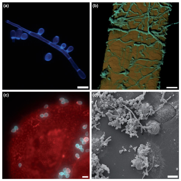
Background: Millions of humans and animals suffer from superficial infections caused by a group of highly specialized filamentous fungi, the dermatophytes, which exclusively infect keratinized host structures. To provide broad insights into the molecular basis of the pathogenicity-associated traits, we report the first genome sequences of two closely phylogenetically related dermatophytes, Arthroderma benhamiae and Trichophyton verrucosum, both of which induce highly inflammatory infections in humans.

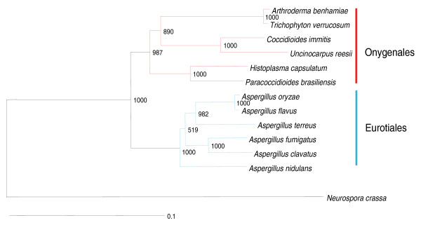
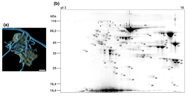
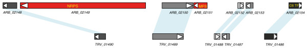
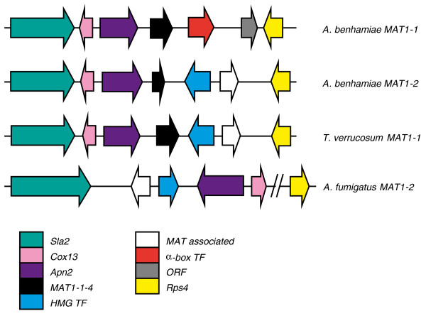
Results: 97% of the 22.5 megabase genome sequences of A. benhamiae and T. verrucosum are unambiguously alignable and collinear. To unravel dermatophyte-specific virulence-associated traits, we compared sets of potentially pathogenicity-associated proteins, such as secreted proteases and enzymes involved in secondary metabolite production, with those of closely related onygenales (Coccidioides species) and the mould Aspergillus fumigatus. The comparisons revealed expansion of several gene families in dermatophytes and disclosed the peculiarities of the dermatophyte secondary metabolite gene sets. Secretion of proteases and other hydrolytic enzymes by A. benhamiae was proven experimentally by a global secretome analysis during keratin degradation. Molecular insights into the interaction of A. benhamiae with human keratinocytes were obtained for the first time by global transcriptome profiling. Given that A. benhamiae is able to undergo mating, a detailed comparison of the genomes further unraveled the genetic basis of sexual reproduction in this species.

Conclusions: Our results enlighten the genetic basis of fundamental and putatively virulence-related traits of dermatophytes, advancing future research on these medically important pathogens.


Figures

Figure 1
Figure 1
Hyphae and microconidia of A. benhamiae on human hair and human keratinocytes. (a) Fluorescence microscopic picture (laser scanning microscope LSM 5 LIVE, Zeiss, Jena) of hyphae and microconidia stained with fluorescent brightener 28 (Sigma, USA). Scale bar: 5 μm. (b) Colonization of human hair. Cyan, fluorescence brightener 28-stained fungal hyphae; orange, hair autofluorescence. Scale bar: 20 μm. (c) Attachment of microconidia to human keratinocytes. Cyan, fluorescence brightener 28-stained fungal hyphae, red, wheat-germ agglutinin stained keratinocytes. Scale bar: 5 μm. (d) Human keratinocytes with germinating A. benhamiae microconidia. Scanning electron microscopy image. Scale bar: 10 μm. See Additional file 1 for supplementary information pertaining to this figure.
Figure 2
Figure 2
Partial genome-based phylogenetic tree of A. benhamiae and T. verrucosum representing the most closely related clades. The tree was inferred by the neighbor-joining analysis method using the PHYLIP package [59], with the number of bootstrap trials set to 1,000. Numbers at the nodes indicate the bootstrap support. See the details and the entire tree in Additional file 3.
Figure 3
Figure 3
Secretome of A. benhamiae grown on keratin. (a) A. benhamiae grown on keratin particles. Cyan, fluorescence brightener 28-stained fungal hyphae; orange, keratin particle autofluorescence. Scale bar: 10 μm. (b) Two-dimensional gel of secreted A. benhamiae proteins obtained from culture supernatant after 48 h cultivation in a shaking flask with 0.9 g/l glucose and 10 g/l keratin. The apparent molecular mass of proteins and the pI range of the first dimension are indicated. Proteins were identified by mass spectrometry (matrix-assisted laser desorption/ionization-time of flight/time of flight (MALDI-TOF/TOF)). Identified proteins are given in Table S5 in Additional file 6. See also Additional file 1 for more details.
Figure 4
Figure 4
A. benhamiae NRPS ARB_02149 gene cluster and the corresponding region in the T. verrucosum genome.
Figure 5
Figure 5
Mating type gene organization of A. benhamiae and T. verrucosum. Genes constituting the MAT locus: Sla2, putative cytoskeleton assembly control protein (ARB_07317, TRV_02048, AFUA_3G06140); Cox13, cytochrome C oxidase subunit VIa (ARB_08059, TRV_08208, AFUA_3G06190); Apn2, DNA lyase (ARB_07318, TRV_02049, AFUA_3G06180); a gene similar to MAT1-1-4 (ARB_07319, TRV_02050); HMG TF, HMG-box transcription factor (MAT1-2-1; ARB_7320, TRV_02051, AFUA_3G06170); MAT associated protein of unknown function (ARB_07321, TRV_02052, AFUA_3G06160); α-box transcription factor (MAT1-1-1, GB GQ996965); ORF, glycine rich protein of unknown function; Rps4, protein S4 of the 40S ribosomal subunit (ARB_7322, TRV_02053, AFUA_3G06840).

References

    1. Weitzman I, Summerbell RC. The dermatophytes. Clin Microbiol Rev. 1995;8:240–259. - PMC - PubMed
    1. Monod M. Secreted proteases from dermatophytes. Mycopathologia. 2008;166:285–294. doi: 10.1007/s11046-008-9105-4. - DOI - PubMed
    1. White TC, Oliver BG, Gräser Y, Henn MR. Generating and testing molecular hypotheses in the dermatophytes. Eukaryot Cell. 2008;7:1238–1245. doi: 10.1128/EC.00100-08. - DOI - PMC - PubMed
    1. Giddey K, Monod M, Barblan J, Potts A, Waridel P, Zaugg C, Quadroni M. Comprehensive analysis of proteins secreted by Trichophyton rubrum and Trichophyton violaceum under in vitro conditions. J Proteome Res. 2007;6:3081–3092. doi: 10.1021/pr070153m. - DOI - PubMed
    1. Liu T, Zhang Q, Wang L, Yu L, Leng W, Yang J, Chen L, Peng J, Ma L, Dong J, Xu X, Xue Y, Zhu Y, Zhang W, Yang L, Li W, Sun L, Wan Z, Ding G, Yu F, Tu K, Qian Z, Li R, Shen Y, Li Y, Jin Q. The use of global transcriptional analysis to reveal the biological and cellular events involved in distinct development phases of Trichophyton rubrum conidial germination. BMC Genomics. 2007;8:100. doi: 10.1186/1471-2164-8-100. - DOI - PMC - PubMed

Publication types

LinkOut - more resources