. 2011 Apr;32(10):2524-31.
doi: 10.1016/j.biomaterials.2010.12.027. Epub 2011 Jan 17.

Hydrogel network design using multifunctional macromers to coordinate tissue maturation in ovarian follicle culture

Affiliations

Hydrogel network design using multifunctional macromers to coordinate tissue maturation in ovarian follicle culture

Ariella Shikanov et al. Biomaterials. 2011 Apr.

Abstract

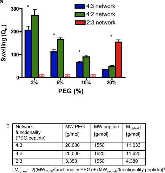
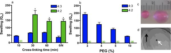
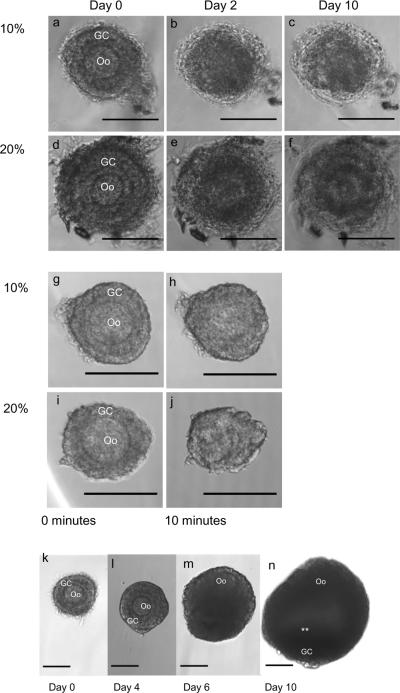
Synthetic hydrogels with tunable properties are appealing for regenerative medicine. A critical limitation in hydrogel design at low solids concentration is the formation of defects, which increase gelation times and swelling, and reduce elasticity. Here, we report that trifunctional cross-linking peptides applied to 4-arm poly-(ethylene glycol) (PEG) hydrogels decreased swelling and gelation time relative to bi-functional crosslinkers. In contrast to bi-functional peptides, the third cross-linking site on the peptide created a branch point if an intramolecular cross-link formed, which prevented non-functional "dangling-ends" in the hydrogel network and enhanced the number of elastically active cross-links. The improved network formation enabled mouse ovarian follicle encapsulation and maturation in vitro. Hydrogels with bi-functional crosslinkers resulted in cellular dehydration, likely due to osmosis during the prolonged gelation. For trifunctional crosslinkers, the hydrogels supported a 17-fold volumetric expansion of the tissue during culture, with expansion dependent on the ability of the follicle to rearrange its microenvironment, which is controlled through the sensitivity of the cross-linking peptide to the proteolytic activity of plasmin. The improved network design enabled ovarian follicle culture in a completely synthetic system, and can advance fertility preservation technology for women facing premature infertility from anticancer therapies.


Figures

Figure 1
Figure 1. Primary loop formation and swelling in end-linked PEG hydrogels
a–d, Cross-linking of a 4-four arm PEG star and a difunctional peptide. (a) Ideally, reactions are intermolecular, which results in elastically active cross-links (blue shading) between PEG macromers, (c) and is favored at high solids content (represented by a grey circle). (b) Primary loop formation leads to elastically inactive cross-links (grey shading), (d) which is favored at low solids content. e–g, Primary loop structure in different networks. (e,f) In the 4:2 and 2:3 network, primary loops will be a “dangling-end” in the network, and thus elastically inactive. (g) In a 4:3 network, if a primary loop forms, a potential branch point is present to continue the network.
Figure 2
Figure 2. Swelling tests of the hydrogels formed from precursors with different functionalities
a, Mass equilibrium swelling (Qm) of hydrogels with networks composed of different macromer functionalities (PEG-Ac : peptide functionality). At all concentrations, a 4:3 network swells the least, but at concentrations in which primary loop formation would be most favored (3%), the difference in swelling is greatest. Cross-linking reactions occurred at the pH with the lowest swelling, in agreement with published conditions[2, 3], to minimize effects of different peptide chemistries. An infinity symbol indicates that a hydrogel either did not form or dissolved during overnight swelling. Error bars are one standard deviation. *Indicates P < 0.001 relative to 4:3 network. (b) Ideal molecular weight between cross-links (Mc,ideal) for the hydrogels. Note that the trifunctional peptide has slightly asymmetric cross-links, which is not accounted for in this model.
Figure 3
Figure 3. Swelling tests under cellular encapsulation conditions that support ovarian follicle viability
a–d Hydrogel cross-linking in an isotonic HEPES buffer at pH 7.4 with PEG-VS. (a) A 4:3 network supported rapid hydrogel formation within 10 minutes, and swelling did not decrease appreciably with an increased cross-linking period, even after overnight (O/N) cross-linking at 37°C. PEG-VS content was 10% w/v. (b) A 10-minute cross-linking period was sufficient for 4:3 hydrogel formation at low PEG content, suggesting that these hydrogels are resilient to defect formation. An infinity sign indicates that a hydrogel did not form or dissolved during swelling. Error bars are one standard deviation (* indicates P<0.001). (c) Swelling of a 4:3 (left) and 4:2 (right) network of 5% PEG cross-linked for 1 hour. Both gels were formed with an initial volume of 20 μl. (d) To encapsulate ovarian follicles (white arrow) 5% PEG-VS hydrogel disks (black arrow) were cast between glass slides and allowed to gel for 5 minutes at 37°C.
Figure 4
Figure 4. Influence of chemical and osmotic stress on follicles during encapsulation and follicle growth in optimized cross-linking conditions
a,f Exposure of ovarian follicles to a triethanolamine (TEA) buffer, rather than HEPES, during encapsulation severely impacts viability. (a,d) Although follicles appear healthy immediately after encapsulation, (b,e) by day 2 the same follicles appear necrotic. (c,f Follicles do not recover from damage induced by TEA (day 10 of culture shown). g–j, Ovarian follicles suspended in (g,h) 10% or (i,j) 20% PEG solution without the cross-linking peptide lose their normal morphology within ten minutes, possibly due to osmotic pressure induced by the PEG. Notably, the membrane of the oocyte (Oo) (h,j) has lost it spherical shape. The extent of morphological change was dependent on the concentration of PEG and time of exposure, suggesting that a low concentration of PEG and a rapid gelation will be more permissive to ovarian follicle encapsulation. k–n, Morphology of a healthy ovarian follicle (k) following encapsulation: a central oocyte (Oo) surrounded by granulosa cells (GC). (l,m) After several days, proliferation of GCs is observed, (n) and after 10 days, follicles have formed an antral cavity in addition to the GC proliferation (white asterisks). The follicles were cultured in YKNS (intermediate degradation rate) condition. Scale bars are 100 μm.
Figure 5
Figure 5. Follicle growth and development is dependent on proteolytic hydrogel degradation
a, Follicles were encapsulated in 5% PEG-VS cross-linked with a peptide with two plasmin degradation sequences (YKNx), and cultured for 10 days. From day 6 to 10, the follicle expansion in the YKNS condition is significantly greater than the YKND and YDKNDR conditions (*, P<0.001). Error bars are SEM. b–e Hydrogels with the YKNR sequence did not maintain the follicle within the hydrogel beyond four days. (b) Follicles cultured in 2D adhere to and migrate on the culture plate, disrupting their 3D shape. (c–e) Follicles after 10 days of culture encapsulated within hydrogels cross-linked with peptides containing (c) YKNS, (d) YKND, and (e) YDKNDR sequences. (f) The YKNS condition had the greatest antral rate formation and volumetric expansion. Different superscripts indicate significant differences (P<0.01). g, Fertilizable eggs arrested at metaphase-II (MII) following IVM of cultured ooytes; egg (e) and polar body (white arrowheads). h, Confocal image of an egg showing a MII spindle from a perpendicular perspective (white arrow); actin (red), DNA (blue), and β-tubulin (green). All scale bars on light micrographs (b–g) are 100 μm and the scale bar on (h) is 25 μm.

References

    1. Lutolf MP, Hubbell J. Synthetic biomaterials as instructive extracellular microenvironments for morphogenesis in tissue engineering. Nat Biotechnol. 2005;23:47–55. - PubMed
    1. Metters A, Hubbell J. Network formation and degradation behavior of hydrogels formed by Michael-type addition reactions. Biomacromolecules. 2005;6:290–301. - PubMed
    1. Lutolf MP, Hubbell JA. Synthesis and physicochemical characterization of end-linked poly(ethylene glycol)-co-peptide hydrogels formed by Michael-type addition. Biomacromolecules. 2003;4:713–22. - PubMed
    1. Pratt AB, Weber FE, Schmoekel HG, Muller R. Synthetic extracellular matrices for in situ tissue engineering. Biotechnol Bioeng. 2004;86:27–36. - PubMed
    1. Rydholm AE, Bowman CN, Anseth KS. Degradable thiol-acrylate photopolymers: polymerization and degradation behavior of an in situ forming biomaterial. Biomaterials. 2005;26:4495–506. - PubMed

Publication types

MeSH terms

Substances