. 2011 May;39(9):3879-91.
doi: 10.1093/nar/gkq1355. Epub 2011 Jan 18.

Identification of human miRNA precursors that resemble box C/D snoRNAs

Affiliations

Identification of human miRNA precursors that resemble box C/D snoRNAs

Motoharu Ono et al. Nucleic Acids Res. 2011 May.

Abstract

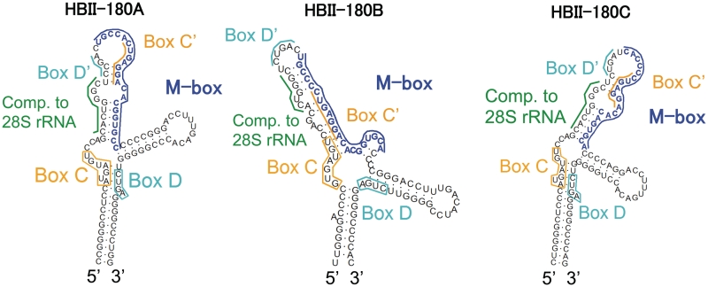
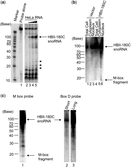
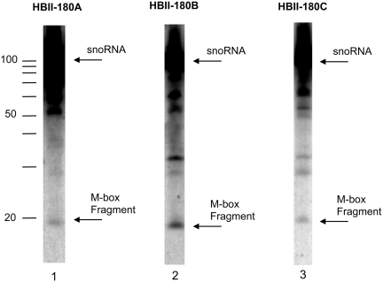
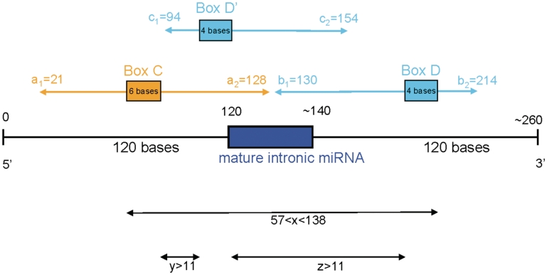
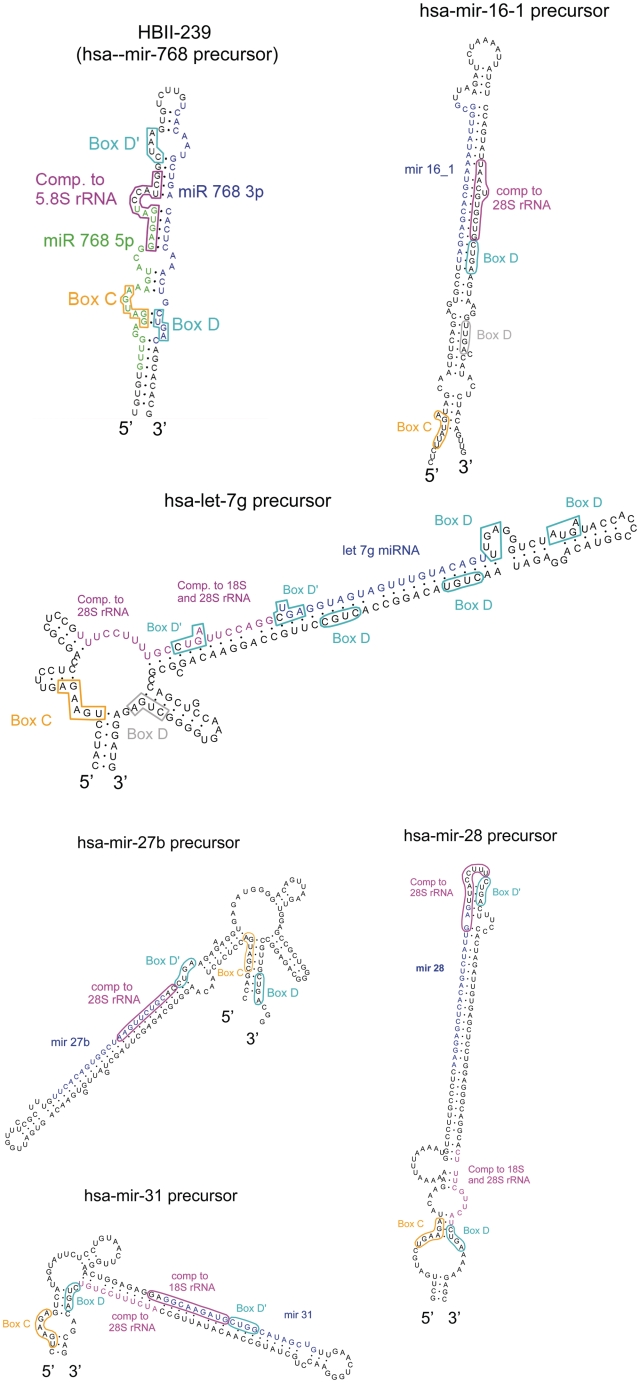
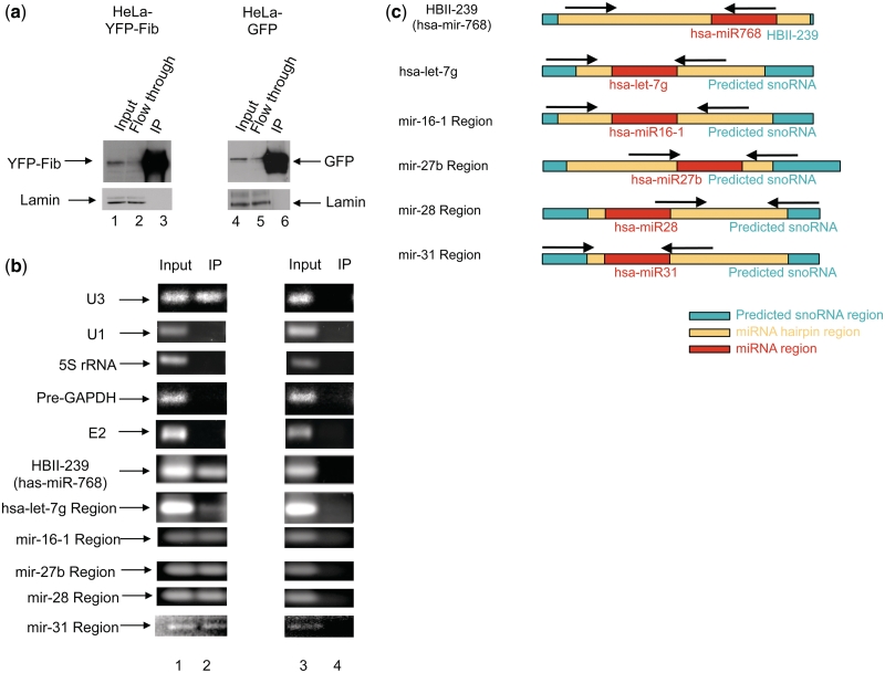
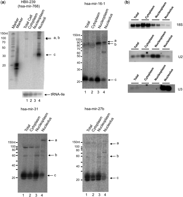
There are two main classes of small nucleolar RNAs (snoRNAs): the box C/D snoRNAs and the box H/ACA snoRNAs that function as guide RNAs to direct sequence-specific modification of rRNA precursors and other nucleolar RNA targets. A previous computational and biochemical analysis revealed a possible evolutionary relationship between miRNA precursors and some box H/ACA snoRNAs. Here, we investigate a similar evolutionary relationship between a subset of miRNA precursors and box C/D snoRNAs. Computational analyses identified 84 intronic miRNAs that are encoded within either box C/D snoRNAs, or in precursors showing similarity to box C/D snoRNAs. Predictions of the folded structures of these box C/D snoRNA-like miRNA precursors resemble the structures of known box C/D snoRNAs, with the boxes C and D often in close proximity in the folded molecule. All five box C/D snoRNA-like miRNA precursors tested (miR-27b, miR-16-1, mir-28, miR-31 and let-7g) bind to fibrillarin, a specific protein component of functional box C/D snoRNP complexes. The data suggest that a subset of small regulatory RNAs may have evolved from box C/D snoRNAs.


Figures

Figure 1.
Figure 1.
Predicted secondary structure of the human HBII-180 box C/D snoRNAs. The characteristic features of members of the box C/D HBII-180 family are shown. Conserved C and D motifs are indicated by orange and cyan boxes/lines, respectively. The positions of rRNA complementary sequence are indicated by a green bar. The positions of M-box regions which can be altered to modulate target gene expression (snoMEN vector) (22) are indicated by a blue bar.
Figure 2.
Figure 2.
Generation of M-box RNA Fragments from HBII-180 snoRNAs. (a) Detection of endogenous HBII-180C M-box fragment by RNase A/T1 protection assay. As a control, the diluted anti-sense probe against HBII-180C was loaded without RNase digestion (Probe lane). The probes were incubated with different amounts of HeLa cell total RNA (0, 1, 5, 10 µg for each, lanes 2–5). Both the mature HBII-180C snoRNA (arrow) and shorter fragments (arrow heads) were protected from RNase A/T1 digestion. (b) Detection of exogenously expressed HBII-180C M-box fragment by northern blotting. The same amount of HeLa cell cytoplasmic RNA (lanes 2 and 5), total RNA (lanes 1 and 4) and nuclear RNA (lanes 3 and 6) were compared with either a transiently transfected HBII-180C expression mini-gene or empty vector (lanes 1–3: Vector, lanes 4–6: HBII-180C). (c) Detection of endogenous HBII-180C M-box fragment by high sensitivity northern blotting. Fractionated HeLa cell nucleolar RNA was blotted on a nylon membrane and HBII-180C M-box fragment detected by hybridization with a radio-labelled RNA oligonucleotide probe (lane 1). The same filter was re-probed using a probe to the 3′ region (box D probe) of snoRNA HBII-180C. The result showed that 3′ region probe detected HBII-180C snoRNA (lane 2) but not any M-box fragment bands even after long exposure time (lane 3).
Figure 3.
Figure 3.
Detection of endogenous M-box RNA fragments of HBII-180 snoRNAs. Equivalent amounts of total HeLa cell RNA were analysed using oligonucleotide probes specific for the respective M-box regions (lanes 1–3).
Figure 4.
Figure 4.
Detailed analysis of HBII-180C M-box fragments. (a) Equivalent amounts of total HeLa cell RNA (i), cytoplasmic RNA (ii), nucleoplasmic RNA (iii) and nucleolar RNA (iv) were separated by PAGE and transferred to a membrane using chemical cross-linking to facilitate high-sensitivity RNA detection as described. HBII-180C M-box RNA fragment sequences were detected using radiolabelled RNA oligonucleotide probes. The relative signal levels that were calculated by imaging software (ImageGauge v4.21, FUJI Photo Film co. Ltd.) are indicated graphically below each blot. The signal-back ground ratio between each fraction was normalized to set the highest signal at 100%. (b) The tRNA sequences were detected using radiolabelled RNA oligonucleotide probes as in (a).
Figure 5.
Figure 5.
Positional characterization of the snoRNA–miRNA molecules. Ten positional parameters (a1, a2, b1, b2, c1, c2, xmin, xmax, y and z) were chosen to describe the position of snoRNA features with respect to the position of the mature miRNA. The values learnt by the genetic algorithm are indicated.
Figure 6.
Figure 6.
Secondary-structure prediction of the HBII-239 box C/D snoRNA which contains the reported miR-768 miRNAs and predicted box C/D snoRNAs encoding known miRNAs. Mature miRNAs are drawn in blue and green. C and D boxes are shown, respectively, in orange and cyan while guide regions are shown in pink. Extra box D motifs not detected by our algorithm are shown in grey.
Figure 7.
Figure 7.
Co-immunoprecipitation of miRNA precursors with fibrillarin. (a) Western blot confirming specificity of the immunoprecipitation using an anti-GFP antibody. Nuclear extracts were prepared from HeLa cells stably expressing either free GFP or YFP-Fibrillarin (YFP-Fib) and immunoprecipitated using an anti-GFP antibody as previously described (22,27). The same membrane was reprobed with an antibody against lamin as a loading control. (b) RT–PCR used to detect co-precipitated HBII-239, hsa-mir-let-7g, hsa-mir-16-1, hsa-mir-27b, has-mir-28 and has-mir-31 miRNA precursors, with U3 box C/D snoRNA as positive control and, U1 snRNA, 5 S rRNA, GAPDH pre-mRNA and E2 box H/ACA snoRNA as negative controls for fibrillarin-associated RNAs. (c) Position of the primers used to detect the specified miRNA extended regions is shown by arrows.
Figure 8.
Figure 8.
Subcellular localization of box C/D snoRNA-like miRNA precursors. (a) Northern blots of HeLa cell extracts fractionated into cytoplasmic, nucleoplasmic and nucleolar fractions were probed for the presence of HBII-239, mir-16-1, mir-27b and mir-31 encoding molecules. In all panels, bands labelled with ‘a’ represent the expected size of the predicted snoRNAs, those labelled with ‘b’ represent the expected size for the miRNA hairpins and ‘c’ represents the expected size of the mature miRNA. (b) The fractionation was further controlled using U3 snoRNA, U2 snRNA and 18S rRNA. Blots show the results of two independent fractionations.

References

    1. Lai EC. microRNAs: runts of the genome assert themselves. Curr. Biol. 2003;13:R925–R936. - PubMed
    1. Baskerville S, Bartel DP. Microarray profiling of microRNAs reveals frequent coexpression with neighboring miRNAs and host genes. RNA. 2005;11:241–247. - PMC - PubMed
    1. Kim YK, Kim VN. Processing of intronic microRNAs. EMBO J. 2007;26:775–783. - PMC - PubMed
    1. Rodriguez A, Griffiths-Jones S, Ashurst JL, Bradley A. Identification of mammalian microRNA host genes and transcription units. Genome Res. 2004;14:1902–1910. - PMC - PubMed
    1. Lai EC. miRNAs: whys and wherefores of miRNA-mediated regulation. Curr. Biol. 2005;15:R458–R460. - PubMed

Publication types