. 2011 May;39(9):3852-63.
doi: 10.1093/nar/gkq1323. Epub 2011 Jan 18.

Mutant CAG repeats of Huntingtin transcript fold into hairpins, form nuclear foci and are targets for RNA interference

Affiliations

Mutant CAG repeats of Huntingtin transcript fold into hairpins, form nuclear foci and are targets for RNA interference

Mateusz de Mezer et al. Nucleic Acids Res. 2011 May.

Abstract

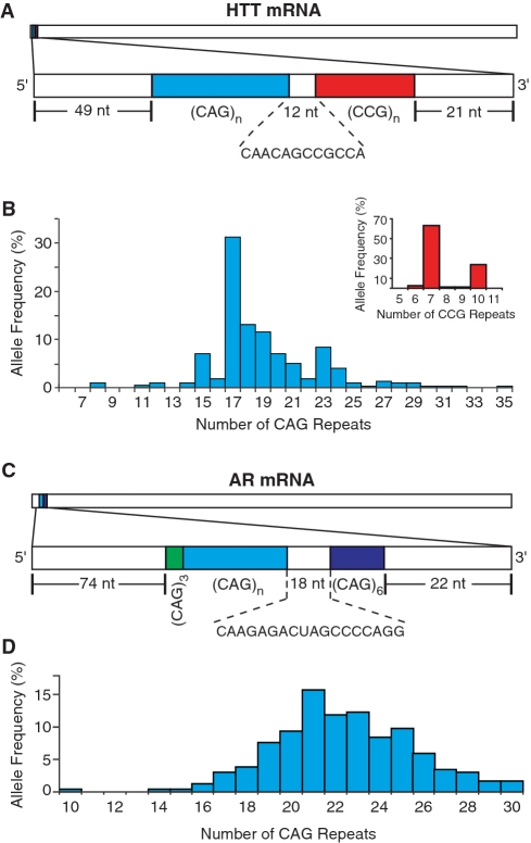
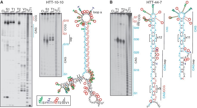
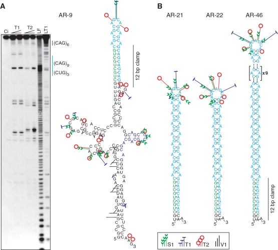
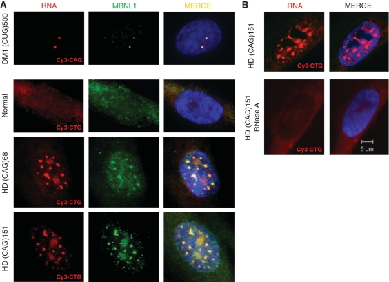
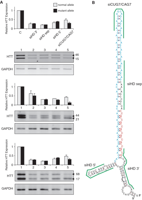
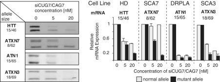
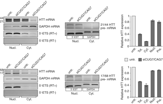
The CAG repeat expansions that occur in translated regions of specific genes can cause human genetic disorders known as polyglutamine (poly-Q)-triggered diseases. Huntington's disease and spinobulbar muscular atrophy (SBMA) are examples of these diseases in which underlying mutations are localized near other trinucleotide repeats in the huntingtin (HTT) and androgen receptor (AR) genes, respectively. Mutant proteins that contain expanded polyglutamine tracts are well-known triggers of pathogenesis in poly-Q diseases, but a toxic role for mutant transcripts has also been proposed. To gain insight into the structural features of complex triplet repeats of HTT and AR transcripts, we determined their structures in vitro and showed the contribution of neighboring repeats to CAG repeat hairpin formation. We also demonstrated that the expanded transcript is retained in the nucleus of human HD fibroblasts and is colocalized with the MBNL1 protein. This suggests that the CAG repeats in the HTT mRNA adopt ds-like RNA conformations in vivo. The intracellular structure of the CAG repeat region of mutant HTT transcripts was not sufficiently stable to be protected from cleavage by an siRNA targeting the repeats and the silencing efficiency was higher for the mutant transcript than for its normal counterpart.


Figures

Figure 1.
Figure 1.
Characteristics of repeat regions from HTT and AR mRNAs. (A and C) Schematic representation of HTT and AR mRNA fragments. White bars, specific flanking sequences; blue bars, polymorphic CAG repeats; red bar, polymorphic CCG repeat region; dark blue bar, monomorphic (CAG)6 tract; green bar, monomorphic (CUG)3 sequence. Genotyping of CAG and CCG repeat tracts in the HTT gene (B) and the polymorphic CAG tract from the AR gene (D).
Figure 2.
Figure 2.
In vitro structure of CAG and CCG repeat tracts from HTT mRNA. (A) Structure analysis of the HTT-10-10 mRNA fragment. The left autoradiogram (short run) represents the pattern of RNA fragments generated by the nucleases: S1 (2.5 u/µl and 5 u/µl), T1 (0.1 u/µl and 0.15 u/µl), T2 (0.1 u/µl and 0.2 u/µl) and V1 (0.1 u/µl and 0.2 u/µl). The middle autoradiogram (medium run) depicts cleavage products generated by S1 nuclease and T1 RNase in the repeat region. Positions of selected G residues from the repeated sequences (G1 shows the G residue of the first triplet repeat) and the 12-nt repeat separation region (sep) are indicated. The right section shows the secondary structure formed by the HTT mRNA fragment. (B) Secondary structure model of the trinucleotide repeat region in the HTT mRNA fragments containing expanded CAG repeats (HTT-44-7). The autoradiogram shows cleavages generated by RNase T1 (0.1 u/µl and 0.15 u/µl) and T2 (0.1 u/µl and 0.2 u/µl) in the repeat region. The middle section presents a model of the secondary structure of the trinucleotide repeat region from HTT-44-7 whereas the right section shows an alternative model of the secondary structure in the terminal loop and CAG/sep region. Structural motifs that are repeated several times are surrounded by brackets. LF and LT1 indicate the 1-nt and G-specific ladders, respectively; Ci, incubation control in which RNA was incubated in the absence of nucleases. Sites and intensity of cleavages induced by the different probes are indicated (inset legend).
Figure 3.
Figure 3.
In vitro structure analysis of repeat region from AR mRNA fragments. (A) The autoradiogram (right section) depicts RNA fragments generated by T1 (0.1 u/µl and 0.15 u/µl) and T2 (0.1 u/µl and 0.2 u/µl) enzymatic probes in the AR-9 transcript (containing 9 CAG repeats). Other symbols are as in the legend to Figure 2. The right section shows the secondary structure formed by the AR-9 transcript. (B) Secondary structures of the trinucleotide repeat region from AR transcripts harboring different numbers of CAG repeats (from left to right, 21, 22 and 46 CAG repeats). The hairpin structure stabilizing interaction between CUG and CAG repeats is indicated by a 12-bp clamp.
Figure 4.
Figure 4.
Mutant CAG RNA repeats form nuclear aggregates in human HD cells and colocalize with MBNL1. (A) Representative images of RNA FISH and IF performed on cultured human fibroblasts. HD cells expressing 68 and 151 CAG repeats from the HTT gene show nuclear accumulation of CAG RNA aggregates, as detected with a CTG probe. As a positive control, human DM1 fibroblasts were included in the FISH and IF assays to detect ribonuclear CUG inclusions with a CAG probe and to show their colocalization with MBNL1 protein. In normal cells, MBNL1 is detected over the entire cell, whereas the presence of CAG RNA foci in HD cells causes colocalization with the splicing factor as depicted also in DM1 cells. CUG and CAG RNA foci are displayed in red, MBNL1 is shown in green and nuclear DNA was stained with DAPI (blue). (B) HD cells expressing 151 CAG repeats were treated with RNase A followed by FISH with the CTG probe. The CAG RNA nuclear aggregates were sensitive to RNase A treatment. CAG RNA foci (red), DAPI nuclear stain (blue).
Figure 5.
Figure 5.
Silencing effects of siRNAs on HTT mRNA expression levels. (A) Results of quantitative RT–PCR showing HTT mRNA expression levels in three human HD cell lines harboring 15/46, 21/44 and 17/68 CAG repeats (CAG repeat sizes from normal and mutant alleles are shown on the side). The cells were either treated with transfection reagent only (lane 1) or treated with one of four indicated siRNAs used at 20 nM (lanes 2–5). Bar graphs represent mean values from three independent experiments normalized to GAPDH expression (SDs are shown as error bars). (B) Potential localization of target sequences (green lines) for the indicated siRNAs on a structural model of the HTT transcript. For siCUG7/CAG7 only one possible binding site is shown.
Figure 6.
Figure 6.
Effect of CUG7/CAG7 siRNA on the silencing of HTT, ATXN7, ATXN3 and ATN1 transcripts. Quantitative RT–PCR analysis of four transcripts containing CAG repeats in human HD, SCA7, SCA3 and DRPLA fibroblasts treated with two different concentrations of CUG7/CAG7 siRNA (left panel). Upper bands represent mutant and lower bands represent normal mRNA variants. Bar graphs (right panel) show relative expression levels of the indicated mRNA variants calculated in three independent experiments and normalized to GAPDH expression (error bars represent SDs).
Figure 7.
Figure 7.
siRNA targeting CAG repeats acts predominantly in the cytoplasm. RT–PCR results for the nuclear and cytoplasmic RNA isolated from HD fibroblasts untransfected and siRNA transfected. The experiments were carried out in triplicate and representative gels are shown. Quantitative results of RT–PCR shown in the bar diagrams represent the relative levels of HTT RNA expression (±SD) normalized to HTT RNA level in untransfected HD cells and either to GAPDH expression (for total and cytoplasmic RNA) or to the 5′-ETS (for the nuclear RNA). As shown, HTT RNA silencing was significantly stronger in the cytoplasm than in the nucleus. Low level of nuclear silencing was confirmed by analysis of HTT pre-mRNA. (RT+) indicates the presence and (RT–) the absence of reverse transcriptase in the reaction; Untr., untransfected; siRNA, siCUG7/CAG7; Nucl., nuclear; Cyt., cytoplasmic; Tot., total; Pre., pre-mRNA.

Similar articles

Cited by

References

    1. Bates GP. History of genetic disease: the molecular genetics of Huntington disease - a history. Nat. Rev. Genet. 2005;6:766–773. - PubMed
    1. Martin JB, Gusella JF. Huntington's disease. Pathogenesis and management. N. Engl. J. Med. 1986;315:1267–1276. - PubMed
    1. The Huntington's Disease Collaborative Research Group. A novel gene containing a trinucleotide repeat that is expanded and unstable on Huntington's disease chromosomes. Cell. 1993;72:971–983. - PubMed
    1. La Spada AR, Wilson EM, Lubahn DB, Harding AE, Fischbeck KH. Androgen receptor gene mutations in X-linked spinal and bulbar muscular atrophy. Nature. 1991;352:77–79. - PubMed
    1. Gatchel JR, Zoghbi HY. Diseases of unstable repeat expansion: mechanisms and common principles. Nat. Rev. Genet. 2005;6:743–755. - PubMed

Publication types