. 2011 Mar 25;286(12):10258-64.
doi: 10.1074/jbc.M110.163436. Epub 2011 Jan 19.

The heat shock-binding protein (HspBP1) protects cells against the cytotoxic action of the Tag7-Hsp70 complex

Affiliations

The heat shock-binding protein (HspBP1) protects cells against the cytotoxic action of the Tag7-Hsp70 complex

Denis V Yashin et al. J Biol Chem. .

Abstract

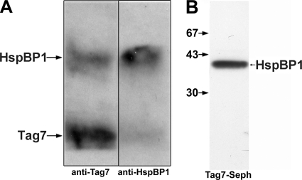
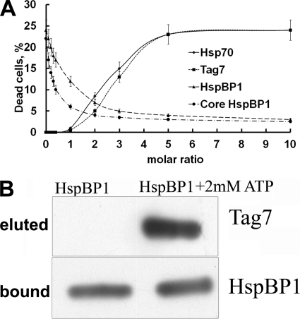
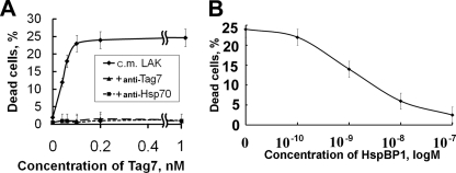
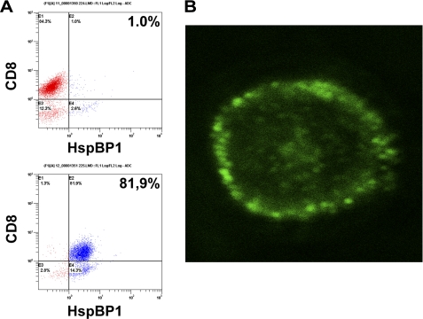
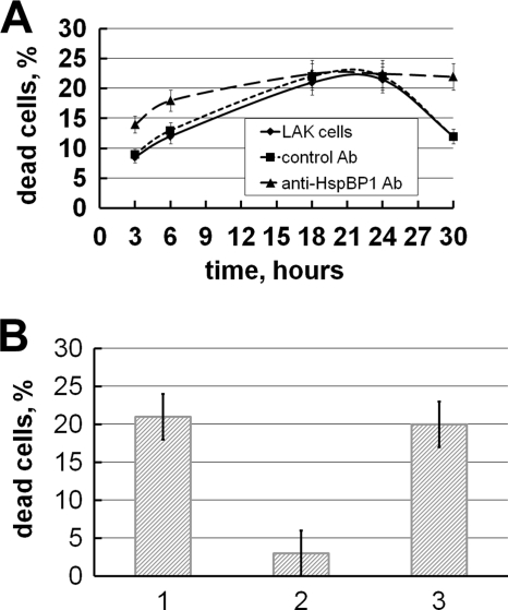
Heat shock-binding protein HspBP1 is a member of the Hsp70 co-chaperone family. The interaction between HspBP1 and the ATPase domain of the major heat shock protein Hsp70 up-regulates nucleotide exchange and reduces the affinity between Hsp70 and the peptide in its peptide-binding site. Previously we have shown that Tag7 (also known as peptidoglycan recognition protein PGRP-S), an innate immunity protein, interacts with Hsp70 to form a stable Tag7-Hsp70 complex with cytotoxic activity against some tumor cell lines. This complex can be produced in cytotoxic lymphocytes and released during interaction with tumor cells. Here the effect of HspBP1 on the cytotoxic activity of the Tag7-Hsp70 complex was examined. HspBP1 could bind not only to Hsp70, but also to Tag7. This interaction eliminated the cytotoxic activity of Tag7-Hsp70 complex and decreased the ATP concentration required to dissociate Tag7 from the peptide-binding site of Hsp70. Moreover, HspBP1 inhibited the cytotoxic activity of the Tag7-Hsp70 complex secreted by lymphocytes. HspBP1 was detected in cytotoxic CD8+ lymphocytes. This protein was released simultaneously with Tag7-Hsp70 during interaction of these lymphocytes with tumor cells. The simultaneous secretion of the cytotoxic complex with its inhibitor could be a mechanism protecting normal cells from the cytotoxic effect of this complex.


Figures

FIGURE 1.
FIGURE 1.
HspBP1 interacts with Tag7. A, Tag7 and HspBP1 were incubated for 30 min in PBS at 10−9 m at room temperature, then applied to anti-Tag7 and anti-HspBP1-Sepharose columns, and washed with PBS plus 0.5 m NaCl, with PBS, and water, followed by elution with 0.25 M TEA (pH 12.5). All eluates were dried, dissolved in a minimal volume of the SDS-PAGE sample buffer, and analyzed on 12% gels. After blotting onto nitrocellulose, the proteins were visualized using anti-Tag7 and anti-HspBP1 antibodies followed by either anti-mouse (Tag-7) or anti-sheep (HspBP1) antibodies and ECL Plus. B, HspBP1 was applied to Tag7-Sepharose, following the same procedures as in A. Western blot was visualized using anti-HspBP1 antibodies and ECL Plus.
FIGURE 2.
FIGURE 2.
HspBP1 inactivates the Tag7-Hsp70 complex. A, cytotoxic assay. Protein pairs [Tag7+HspBP1 (♦), Hsp70+HspBP1 (■), Tag7+Hsp70 (▴), Tag7+Hsp70 (●)] were incubated at equal molar concentrations (10−10 m) for 30 min at 20 °C under sterile conditions. Increasing amounts of the third protein were added to each pair, Hsp70 (♦), Tag7 (■), HspBP1(▴), or core fragment of HspBP1 (●). The mixtures was incubated for another 30 min and assayed for cytoxicity against L929 cells. The mean percentage of cell killing (± S.E., n = 5) by the protein mixture (final dilution 1:10) is plotted against increasing concentrations of the third added protein. B, dissociation of Tag7 from Tag7-Hsp70 complex by HspBP1. Tag7-Hsp70 complex was first applied to anti-Tag7-Sepharose to remove the free Hsp70. The bound complex was eluted with 0.25 M TEA (pH 12.5), dialyzed against PBS (pH 7.5) and applied to anti-Hsp70 Sepharose. The column was washed with PBS containing 0.5 m NaCl, with PBS, and H2O. HspBP1 or HspBP1 with 2 mm ATP in PBS were applied to the loaded material (upper lane indicates eluted proteins). Proteins remaining on the column were eluted with 0.25 M TEA (pH 12.5), separated by SDS-PAGE, blotted onto nitrocellulose, and visualized using anti-Tag7 and anti-HspBP1 antibodies as described under “Experimental Procedures.”
FIGURE 3.
FIGURE 3.
Inhibition of LAK-released cytotoxic activity. A, cytotoxic activity of conditioned media from LAK cells (♦), with 100 nm anti-Tag7 (▴) or anti-Hsp70 antibodies (■). Conditioned media were concentrated using Centricons (Millipore) and dissolved by adding RPMI medium to achieve a needed concentration of Tag7. B, inhibition of cytotoxic activity of conditioned medium from LAK cells by addition of recombinant HspBP1.
FIGURE 4.
FIGURE 4.
HspBP1 is located under the lymphocyte surface. A, HspBP1 on the surface of LAK cells (top) and inside the cell (bottom) as determined by flow cytometry. B, confocal image of a permeabilized CD8+ cell stained with anti-HspBP1 antibodies. (10.0×/1.40 NA oil objective).
FIGURE 5.
FIGURE 5.
HspBP1 antibody restores the cytotoxity of LAK cell supernatant. A, time-dependent cytotoxic activity of conditioned medium from LAK cells after incubation with K562 cells, (♦) conditioned medium alone, (▴) incubation with 100 nm anti-HspBP1 antibodies, and (■) incubation with 100 nm preimmune IgG. B, reactivation of cytoxicity of conditioned medium after 36 h. 1, Initial activity of conditioned medium of LAK cells; 2, activity after 30 h at +4 °C; 3, activity after 30 h in the presence of 100 nm anti-HspBP1 antibodies.

References

    1. Bukau B., Deuerling E., Pfund C., Craig E. A. (2000) Cell. 101, 119–122 - PubMed
    1. Bukau B., Weissman J., Horwich A. (2006) Cell. 125, 443–451 - PubMed
    1. DeLuca-Flaherty C., McKay D. B., Parham P., Hill B. L. (1990) Cell 62, 875–887 - PubMed
    1. Bukau B., Horwich A. L. (1998) Cell. 92, 351–366 - PubMed
    1. Greene L. E., Zinner R., Naficy S., Eisenberg E. (1995) J. Biol. Chem. 270, 2967–2973 - PubMed

Publication types

MeSH terms