. 2011 Mar 25;286(12):10238-47.
doi: 10.1074/jbc.M110.205153. Epub 2011 Jan 19.

The dynamics and mechanism of SUMO chain deconjugation by SUMO-specific proteases

Affiliations

The dynamics and mechanism of SUMO chain deconjugation by SUMO-specific proteases

Miklós Békés et al. J Biol Chem. .

Abstract

SUMOylation of proteins is a cyclic process that requires both conjugation and deconjugation of SUMO moieties. Besides modification by a single SUMO, SUMO chains have also been observed, yet the dynamics of SUMO conjugation/deconjugation remain poorly understood. Using a non-deconjugatable form of SUMO we demonstrate the underappreciated existence of SUMO chains in vivo, we highlight the importance of SUMO deconjugation, and we demonstrate the highly dynamic nature of the SUMO system. We show that SUMO-specific proteases (SENPs) play a crucial role in the dynamics of SUMO chains in vivo by constant deconjugation. Preventing deSUMOylation in Schizosaccharomyces pombe results in slow growth and a sensitivity to replication stress, highlighting the biological requirement for deSUMOylation dynamics. Furthermore, we present the mechanism of SUMO chain deconjugation by SENPs, which occurs via a stochastic mechanism, resulting in cleavage anywhere within a chain. Our results offer mechanistic insights into the workings of deSUMOylating proteases and highlight their importance in the homeostasis of (poly)SUMO-modified substrates.


Figures
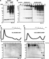
FIGURE 1.
FIGURE 1.
SUMO2 with a P4 mutation from Gln to Pro (Q90P) is not deconjugated by endogenous SENPs leading to a shift in deSUMOylation dynamics. A, HA-tagged wild-type and Q90P SUMO2 were transfected into HEK293A cells and lysed in the presence or absence of 30 mm NEM after 48 h. Lysates were separated by SDS-PAGE and visualized by Western blotting for the HA tag, demonstrating cleavage resistance of the Q90P mutant. B, lysates (+NEM) from time course expression analyses of HEK293A cells transfected with either HA-tagged wild-type or Q90P SUMO2 constructs were separated by SDS-PAGE and visualized by Western blotting for the HA tag, revealing the differential conjugation extent of the Q90P mutant. Dotted line indicates cropping within the same gel. HEK293A cells expressing either HA-SUMO2-WT (C) or HA-SUMO2-Q90P (D) were lysed in the presence of 30 mm NEM, and the lysates were separated on a Superose-6 size exclusion column. UV-trace of lysates is shown on the top and Western blot for the HA tag of the fractions indicated is shown on the bottom, displaying a chain-like banding pattern of SUMO conjugates.
FIGURE 2.
FIGURE 2.
SUMO2 conjugates in the Q90P SUMO2 HMW fractions consist of polySUMO2 chains. A, HMW fractions from lysates of HEK293A cells expressing wild-type (left panel) or Q90P mutant (right panel) SUMO2 were obtained by size exclusion chromatography and the fractions were treated with 3-fold serial dilutions of 1 μm ΔN-SENP1, separated by SDS-PAGE and visualized by Western-blotting for the HA tag. Multimeric cleavage intermediates are indicated on the right. B, time course cleavage assay of HMW-conjugates of wt and Q90P SUMO2 with 1 μm ΔN-SENP1. C, quantification of cleavage of wild-type and Q90P HMW-conjugates from a duplicate experiment (as in A) demonstrates that the Q90P SUMO2 is eventually cleaved, but with a 1000-fold increase in EC50. A parallel experiment using ΔN-SENP1 to cleave HMW-conjugates from N-all-Arg-SUMO2-Q90P expressing cells is shown in D, notice the absence of multimeric cleavage intermediates.
FIGURE 3.
FIGURE 3.
Cleavage of E. coli-produced polySUMO2 chains. A, lysates producing SUMO2 chains were prepared from E. coli (see “Experimental Procedures”) and were treated with 100 nm ΔN-SENP1 for 1 h at 37 °C to show complete cleavage of SUMO2 chains. Coomassie-stained total lysates are shown on the left, and the corresponding Western blot for SUMO2/3 on the right. The asterisks indicate presumed nonspecific bands. B, total E. coli lysate expressing SUMO2 chains was subjected to trypsin digest and LC-MS/MS and searched using Sequest and SUMmOn algorithms. The top panel shows the total ion current, in the middle the distribution of the parent ion shown for the identified K11-linked SUMO-SUMO adduct and the bottom panel shows the distribution of the parent ion for the K42-linked SUMO-SUMO adduct. Purified SUMO2 chains (C) or purified polySUMO2-RanGAP1-C2 (D) were cleaved with equal serial dilutions of ΔN-SENP1 and ΔN-SENP6 for 30 min at 37 °C then separated by SDS-PAGE and visualized by Western blotting for SUMO2/3 or stained with Coomassie stain, demonstrating the extent of SUMO2 chain deconjugation.
FIGURE 4.
FIGURE 4.
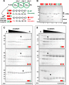
Linear tri-SUMO2 constructs are cleaved in a stochastic manner by ΔN-SENP1 and by ΔN-SENP6. A, schematic representation of linear tri-SUMO2 constructs. Open green circles represent cleavable (WT) ΔN11-SUMO2 and full red circles represent uncleavable (Q90P) ΔN11-SUMO2. The lighting bolt indicates the cleavage site. B, purified tri-SUMO2 proteins were diluted to 1 μm and cleaved with 50 nm ΔN-SENP1 for 30 min at 37 °C and visualized by SDS-PAGE and Coomassie Blue staining. All tri-SUMO2 proteins are only cleaved at the allowed wild-type cleavage site (where P4 is Gln) and not when P4 is mutated to Pro (Q90P). In panels C and D the different tri-SUMO2 proteins (2 μm) were cleaved with 2-fold serial dilutions of 1 μm ΔN-SENP1 (C) or ΔN-SENP6 (D) for 30 min at 37 °C and visualized by SDS-PAGE and Coomassie Blue staining. The order of cleavage sites within tri-SUMO2 for each panel is indicated in the bottom right corner by the order of the red and green circles. E stands for lane with enzyme alone, and S stands for lane with substrate only. Panels in D have been cropped for clarity. The black triangles (▾) indicate approximate EC50 values.
FIGURE 5.
FIGURE 5.
In vivo consequences of the lack of deSUMOylation. A, Western blot analysis of the indicated S. pombe strains was performed using denaturing lysis conditions to analyze total levels of SUMOylated proteins in vivo. Cells were grown in minimal media, that either permitted (−B1, left panel), or repressed gene expression (+B1, right panel). The asterisk indicates a nonspecific band. B, 5-fold dilutions of the indicated strains were grown in minimal media, which either repressed (+B1), or de-repressed expression (−B1) of the indicated genes, in either the presence, or absence of the replication inhibitor hydroxyurea. C, growth curve of double knocked-down and reconstituted HEK293A cells. Cells were grown in 24-well plates and counted every day for 3 days post-transfection by Trypan Blue staining (the error bars represent S.E. of the experiment in duplicate). D, Western blot analysis of the cells shown in panel E. ns and S stand for nonspecific and SUMO-2 + 3 siRNA, respectively. Asterisks (*) indicate nonspecific bands.
FIGURE 6.
FIGURE 6.
Modeling isopeptide-linked SUMO2 chains. Modeling a 5-mer isopeptide-linked SUMO2 chain based on the RanGAP-SUMO linkage from the crystal structure of RanGAP1-SUMO1:SENP2 (PDB: 2IO2) (45) shows how the protease (in ribbon structure with transparent surface in pink and light brown) has sufficient binding capability at any given SUMO2-SUMO2 isopeptide linkage (surface representation of linked SUMO2 molecules in shades of blue). The image was generated using Pymol.

References

    1. Schwartz D. C., Hochstrasser M. (2003) Trends Biochem. Sci. 28, 321–328 - PubMed
    1. Hershko A. (1983) Cell 34, 11–12 - PubMed
    1. Tatham M. H., Jaffray E., Vaughan O. A., Desterro J. M., Botting C. H., Naismith J. H., Hay R. T. (2001) J. Biol. Chem. 276, 35368–35374 - PubMed
    1. Knipscheer P., van Dijk W. J., Olsen J. V., Mann M., Sixma T. K. (2007) EMBO J. 26, 2797–2807 - PMC - PubMed
    1. Matic I., van Hagen M., Schimmel J., Macek B., Ogg S. C., Tatham M. H., Hay R. T., Lamond A. I., Mann M., Vertegaal A. C. (2008) Mol. Cell Proteomics 7, 132–144 - PMC - PubMed

Publication types

MeSH terms