

Exploring by pulsed EPR the electronic structure of ubisemiquinone bound at the QH site of cytochrome bo3 from Escherichia coli with in vivo 13C-labeled methyl and methoxy substituents
- PMID: 21247900
- PMCID: PMC3060462
- DOI: 10.1074/jbc.M110.206821
Exploring by pulsed EPR the electronic structure of ubisemiquinone bound at the QH site of cytochrome bo3 from Escherichia coli with in vivo 13C-labeled methyl and methoxy substituents
Abstract
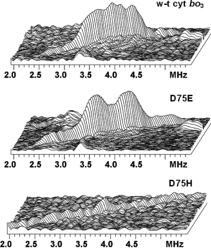
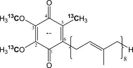
The cytochrome bo(3) ubiquinol oxidase from Escherichia coli resides in the bacterial cytoplasmic membrane and catalyzes the two-electron oxidation of ubiquinol-8 and four-electron reduction of O(2) to water. The one-electron reduced semiquinone forms transiently during the reaction, and the enzyme has been demonstrated to stabilize the semiquinone. The semiquinone is also formed in the D75E mutant, where the mutation has little influence on the catalytic activity, and in the D75H mutant, which is virtually inactive. In this work, wild-type cytochrome bo(3) as well as the D75E and D75H mutant proteins were prepared with ubiquinone-8 (13)C-labeled selectively at the methyl and two methoxy groups. This was accomplished by expressing the proteins in a methionine auxotroph in the presence of l-methionine with the side chain methyl group (13)C-labeled. The (13)C-labeled quinone isolated from cytochrome bo(3) was also used for the generation of model anion radicals in alcohol. Two-dimensional pulsed EPR and ENDOR were used for the study of the (13)C methyl and methoxy hyperfine couplings in the semiquinone generated in the three proteins indicated above and in the model system. The data were used to characterize the transferred unpaired spin densities on the methyl and methoxy substituents and the conformations of the methoxy groups. In the wild type and D75E mutant, the constraints on the configurations of the methoxy side chains are similar, but the D75H mutant appears to have altered methoxy configurations, which could be related to the perturbed electron distribution in the semiquinone and the loss of enzymatic activity.
Figures
Similar articles
-
Characterization of mutants that change the hydrogen bonding of the semiquinone radical at the QH site of the cytochrome bo3 from Escherichia coli.J Biol Chem. 2007 Mar 23;282(12):8777-85. doi: 10.1074/jbc.M611595200. Epub 2007 Jan 30. J Biol Chem. 2007. PMID: 17267395
-
Studies on a stabilisation of ubisemiquinone by Escherichia coli quinol oxidase, cytochrome bo.Eur J Biochem. 1995 Feb 1;227(3):903-8. doi: 10.1111/j.1432-1033.1995.tb20217.x. Eur J Biochem. 1995. PMID: 7867653
-
Identification of the residues involved in stabilization of the semiquinone radical in the high-affinity ubiquinone binding site in cytochrome bo(3) from Escherichia coli by site-directed mutagenesis and EPR spectroscopy.Biochemistry. 2002 Aug 27;41(34):10675-9. doi: 10.1021/bi012146w. Biochemistry. 2002. PMID: 12186553
-
QH*- ubisemiquinone radical in the bo3-type ubiquinol oxidase studied by pulsed electron paramagnetic resonance and hyperfine sublevel correlation spectroscopy.Biochemistry. 2001 Jan 30;40(4):1037-43. doi: 10.1021/bi001641+. Biochemistry. 2001. PMID: 11170426
-
Ubiquinol oxidation in the cytochrome bc1 complex: reaction mechanism and prevention of short-circuiting.Biochim Biophys Acta. 2005 Aug 15;1709(1):5-34. doi: 10.1016/j.bbabio.2005.03.009. Epub 2005 Apr 8. Biochim Biophys Acta. 2005. PMID: 16005845 Review.
Cited by
-
Structure of the cytochrome aa3 -600 heme-copper menaquinol oxidase bound to inhibitor HQNO shows TM0 is part of the quinol binding site.Proc Natl Acad Sci U S A. 2020 Jan 14;117(2):872-876. doi: 10.1073/pnas.1915013117. Epub 2019 Dec 30. Proc Natl Acad Sci U S A. 2020. PMID: 31888984 Free PMC article.
-
Conformational differences between the methoxy groups of QA and QB site ubisemiquinones in bacterial reaction centers: a key role for methoxy group orientation in modulating ubiquinone redox potential.Biochemistry. 2013 Jul 9;52(27):4648-55. doi: 10.1021/bi400489b. Epub 2013 Jun 24. Biochemistry. 2013. PMID: 23745576 Free PMC article.
-
Q-Band Electron-Nuclear Double Resonance Reveals Out-of-Plane Hydrogen Bonds Stabilize an Anionic Ubisemiquinone in Cytochrome bo3 from Escherichia coli.Biochemistry. 2016 Oct 11;55(40):5714-5725. doi: 10.1021/acs.biochem.6b00669. Epub 2016 Sep 28. Biochemistry. 2016. PMID: 27622672 Free PMC article.
-
The semiquinone at the Qi site of the bc1 complex explored using HYSCORE spectroscopy and specific isotopic labeling of ubiquinone in Rhodobacter sphaeroides via (13)C methionine and construction of a methionine auxotroph.Biochemistry. 2014 Sep 30;53(38):6022-31. doi: 10.1021/bi500654y. Epub 2014 Sep 17. Biochemistry. 2014. PMID: 25184535 Free PMC article.
-
Oxygen as Acceptor.EcoSal Plus. 2015;6(2):10.1128/ecosalplus.ESP-0012-2015. doi: 10.1128/ecosalplus.ESP-0012-2015. EcoSal Plus. 2015. PMID: 26734697 Free PMC article. Review.
References
-
- Fisher N., Rich P. R. (2000) J. Mol. Biol. 296, 1153–1162 - PubMed
-
- Lubitz W., Feher G. (1999) Appl. Magn. Reson. 17, 1–48
-
- Abramson J., Riistama S., Larsson G., Jasaitis A., Svensson-Ek M., Laakkonen L., Puustinen A., Iwata S., Wikström M. (2000) Nat. Struct. Biol. 7, 910–917 - PubMed
-
- Crofts A. R. (2004) Annu. Rev. Physiol. 66, 689–733 - PubMed
Publication types
MeSH terms
Substances
Grants and funding
LinkOut - more resources
Full Text Sources
Other Literature Sources
Molecular Biology Databases
