. 2011 Mar;138(5):873-8.
doi: 10.1242/dev.056465. Epub 2011 Jan 19.

Lack of the ventral anterior homeodomain transcription factor VAX1 leads to induction of a second pituitary

Affiliations

Lack of the ventral anterior homeodomain transcription factor VAX1 leads to induction of a second pituitary

Kapil Bharti et al. Development. 2011 Mar.

Abstract

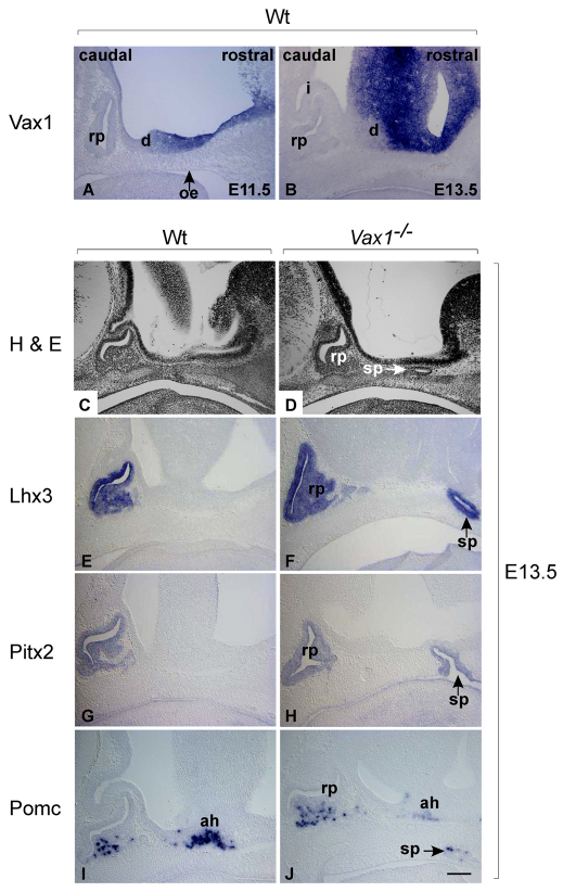
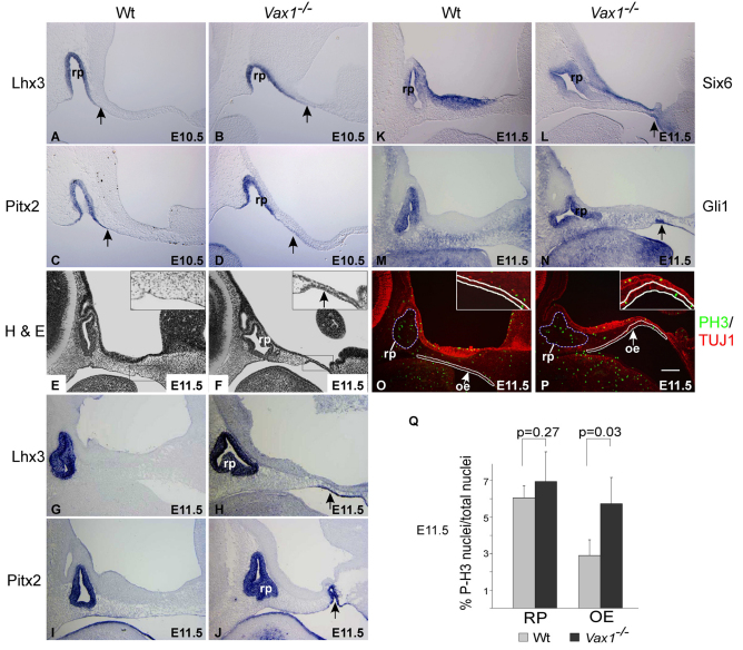
The pituitary gland is an endocrine organ that is developmentally derived from a fold in the oral ectoderm and a juxtaposed fold in the neural ectoderm. Here, we show that the absence of Vax1, a homeodomain transcription factor known for its role in eye and optic chiasm development, causes the rostral oral ectoderm to form an ectopic fold that eventually develops into a separate second pituitary with all the pituitary cell types and neuronal fibers characteristic of the normal pituitary. The induction of the second pituitary is associated with a localized ectopic expression of Fgf10, a gene encoding a growth factor known to recruit oral ectodermal cells into the pituitary. Interestingly, there are rare cases of pituitary duplications in humans that are also associated with optic nerve dysplasia, suggesting that VAX1 might be involved in the pathogenesis of this disorder.


Figures

Fig. 1.
Fig. 1.
Loss of Vax1 leads to induction of a second pouch-like structure in the ventral diencephalon. For all figures, caudal is on the left and rostral is on the right. (A,B) Vax1 in situ hybridization on wild-type sections at the indicated times. In wild type, Vax1 is not seen in oral ectoderm, Rathke's pouch or infundibulum. (C,D) Hematoxylin and Eosin staining of E13.5 wild-type and Vax1−/− sections. Arrow in D shows a distinct, pouch-like structure. (E-J) In situ hybridization on E13.5 Vax1−/− sagittal sections shows ectopic signals (arrows) for the pituitary markers Lhx3 (E,F), Pitx2 (G,H) and Pomc (I,J). ah, anterior hypothalamus; d, diencephalon; i, infundibulum; oe, oral ectoderm; rp, Rathke's pouch; sp, second pouch. Scale bar: 113 μm for A,B,E-J; 141 μm for C,D.
Fig. 2.
Fig. 2.
The second pituitary expresses both anterior and posterior pituitary markers. (A,B) Hematoxylin and Eosin staining of E17.5 wild-type (A) and Vax1−/− (B) sagittal section, showing a large ectopic second pouch, marked with an arrow and arrowhead. (C-H) In situ hybridization for the indicated genes. (I-T) immunostaining for the indicated proteins. (I-L) Co-staining with TUJ1 antibody that labels neurons. For P,T, the second anterior pituitary is marked with arrows and the second posterior pituitary with arrowheads. M and N, O and P, Q and R, and S and T represent paired caudal and rostral parts of the ventral diencephalon. ap, anterior pituitary; pp, posterior pituitary; sp, second pituitary. Scale bar: 42 μm for A,B; 48 μm for C-H; 60 μm for I-L; 110 μm for M-T.
Fig. 3.
Fig. 3.
The second pouch in Vax1 mutants is induced from the rostral oral ectoderm. (A-D) In situ hybridization on E10.5 sections shows a slight rostral expansion of Lhx3 (A,B) and Pitx2 labeling (C,D) in Vax1 mutants (arrows). (E,F) Hematoxylin and Eosin staining of E11.5 wild-type and Vax1−/− sections. Higher magnification in the inset marks thickening in Vax1 mutant rostral oral ectoderm (arrow). (G-N) In situ hybridization performed on sections from E11.5 wild-type and Vax1−/− embryos shows rostral ectopic signals (arrows) for the indicated genes. (O,P) E11.5 embryo sections stained for phosphohistone H3 (green) and TUJ1 (red) show an increased number of phosphohistone H3-positive nuclei in the rostral oral ectoderm (marked with white lines) but not in Rathke's pouch (marked with stippled white lines) of Vax1 mutants. Insets show magnification of rostral oral ectoderm. (Q) Quantitation of number of nuclei positive for phosphohistone H3 labeling, from three mid-sagittal sections from three embryos each. Data are mean±s.d. oe, oral ectoderm; rp, Rathke's pouch. Scale bar: 141 μm for A-D,G-N; 113 μm for E,F,O,P.
Fig. 4.
Fig. 4.
Absence of Vax1 induces Fgf10 expression in the rostral neuroectoderm. (A-D) In situ hybridization for Tcf7L2 (A,B) and Bmp4 (C,D) indicates unchanged expression in the pituitary neuroectoderm (arrowheads). Arrow indicates the second pouch. (E-H) In situ hybridization for Fgf10 shows ectopic expression in the rostral neuroectoderm (arrowhead) at E11.5 (E,F) and E12.5 (G,H). (I,J) Immunohistochemistry for pERK1/2, showing ectopic signal in rostral neuroectoderm (arrowhead) and rostral oral ectoderm (arrows; compare higher magnifications in insets). (K) VAX1 binds to conserved putative homeodomain-binding sites containing subregions of the Fgf10 promoter in vivo. Amplicon positions are indicated relative to the translation start site (see also Fig. S4 in the supplementary material). Amplicons ‘b’ and ‘e’ show positive bands in wild type but not in Vax1−/− mutants. (L) A regulatory model highlighting the role of Vax1 in pituitary induction (for details, see text). Scale bar: 113 μm for A-J.

References

    1. Alstein M., Whitnall M. H., House S., Key S., Gainer H. (1988). An immunochemical analysis of oxytocin and vasopressin prohormone processing in vivo. Peptides 9, 87-105 - PubMed
    1. Bertuzzi S., Hindges R., Mui S. H., O'Leary D. D., Lemke G. (1999). The homeodomain protein vax1 is required for axon guidance and major tract formation in the developing forebrain. Genes Dev. 13, 3092-3105 - PMC - PubMed
    1. Bharti K., Liu W., Csermely T., Bertuzzi S., Arnheiter H. (2008). Alternative promoter use in eye development: the complex role and regulation of the transcription factor MITF. Development 135, 1169-1178 - PMC - PubMed
    1. Brinkmeier M. L., Potok M. A., Davis S. W., Camper S. A. (2007). TCF4 deficiency expands ventral diencephalon signaling and increases induction of pituitary progenitors. Dev. Biol. 311, 396-407 - PMC - PubMed
    1. Charles M. A., Suh H., Hjalt T. A., Drouin J., Camper S. A., Gage P. J. (2005). PITX genes are required for cell survival and Lhx3 activation. Mol. Endocrinol. 19, 1893-1903 - PubMed

Publication types