. 2011 Mar;138(5):811-20.
doi: 10.1242/dev.061416. Epub 2011 Jan 19.

Dynamic stage-specific changes in imprinted differentially methylated regions during early mammalian development and prevalence of non-CpG methylation in oocytes

Affiliations

Dynamic stage-specific changes in imprinted differentially methylated regions during early mammalian development and prevalence of non-CpG methylation in oocytes

Shin-ichi Tomizawa et al. Development. 2011 Mar.

Abstract

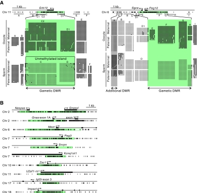
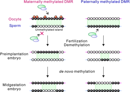
Mammalian imprinted genes are associated with differentially methylated regions (DMRs) that are CpG methylated on one of the two parental chromosomes. In mice, at least 21 DMRs acquire differential methylation in the germline and many of them act as imprint centres. We previously reported the physical extents of differential methylation at 15 DMRs in mouse embryos at 12.5 days postcoitum. To reveal the ontogeny of differential methylation, we determined and compared methylation patterns of the corresponding regions in sperm and oocytes. We found that the extent of the gametic DMRs differs significantly from that of the embryonic DMRs, especially in the case of paternal gametic DMRs. These results suggest that the gametic DMR sequences should be used to extract the features specifying methylation imprint establishment in the germline: from this analysis, we noted that the maternal gametic DMRs appear as unmethylated islands in male germ cells, which suggests a novel component in the mechanism of gamete-specific marking. Analysis of selected DMRs in blastocysts revealed dynamic changes in allelic methylation in early development, indicating that DMRs are not fully protected from the major epigenetic reprogramming events occurring during preimplantation development. Furthermore, we observed non-CpG methylation in oocytes, but not in sperm, which disappeared by the blastocyst stage. Non-CpG methylation was frequently found at maternally methylated DMRs as well as non-DMR regions, suggesting its prevalence in the oocyte genome. These results provide evidence for a unique methylation profile in oocytes and reveal the surprisingly dynamic nature of DMRs in the early embryo.


Figures

Fig. 1.
Fig. 1.
DNA methylation patterns and the extents of differential methylation obtained by bisulphite sequence analysis at the twelve maternally methylated DMRs in oocytes and sperm. (A) Examples of primary bisulphite sequencing results (before the removal of potential clonal amplification events) showing methylated (filled circles) and unmethylated (open circles) CpG sites of two representative DMRs at the mouse Grb10 and Peg10 loci. CpG sites at the DMRs are shown by short vertical lines below the gene organisation schemes; PCR amplicons are indicated by horizontal bars. In the bisulphite sequencing profiles shown, hyphens represent missing or undetermined CpG sites due to SNPs or sequencing problems. Paternal and maternal alleles were discriminated by SNPs if available. The extents of the gametic DMRs determined are shown by the green areas. The Grb10 DMR can be seen as an unmethylated island on the sperm DNA (see text). An additional DMR found at the Peg10 locus is shown in grey. The endpoint of the additional DMR was not determined and therefore the 5′ end is shown by an open box. (B) Extents of the remainder of the maternally methylated DMRs analysed in this study. The DMR in the gametes determined at each locus is shown in green. The precise 3′ endpoint of the Mest DMR was not determined due to repetitive sequences overlapping this region.
Fig. 2.
Fig. 2.
DNA methylation patterns obtained and extents of differential methylation determined at the three paternally methylated DMRs in oocytes and sperm. (A) CpG methylation profile of the mouse H19 (top) and Rasgrf1 (bottom) DMRs in the gametes. Additional DMRs (grey) were found separate from the gametic DMRs containing the imprint centres (ICs, green) at both loci. The gametic and additional DMRs extended over long distances and the endpoints were not determined (denoted by open boxes). Tandem repeat regions are represented by triangles. (B) The extent of the gametic DMR determined at the Dlk1-Gtl2 locus in oocytes and sperm.
Fig. 3.
Fig. 3.
Developmental changes in CpG methylation and DMR extent. DNA methylation status in the gametes (oocyte, red; sperm, blue) compared with that in 12.5 dpc mouse embryos (maternal allele, red; paternal allele, blue). The extents of the DMRs shown were determined after the removal of potential clonal amplification events. Undetermined endpoints of methylation are indicated by gradient shading. Gametic DMRs containing ICs are centred and the changes in DMR extent that have occurred in embryos by 12.5 dpc are represented by pink (maternal DMRs) and light blue (paternal DMRs) areas. Major transcription start sites are indicated by arrows. DMRs were classified into four groups (enlarged, shifted, contracted and fused) according to the pattern of change in extent.
Fig. 4.
Fig. 4.
Developmental changes of DMRs in preimplantation mouse embryos. CpG methylation at three maternally methylated DMRs (Peg10, enlarged DMR; Snrpn, shifted DMR; Impact, contracted DMR) and two paternally methylated DMRs (Dlk1-Gtl2, contracted DMR; H19, fused DMR) were analysed in blastocysts and compared with those in the gametes and 12.5 dpc embryos. CpG sites are indicated by short vertical bars below gene organisations. Horizontal bars indicate positions of PCR amplicons used. Methylation level at each CpG site is shown by vertical bars, with the height of each bar representing the degree of methylation (red, oocyte and maternal allele; blue, sperm and paternal allele). Potential clonal amplification events were removed. Where there was no SNP for allele discrimination, methylation level is indicated by black bars. DMRs are highlighted in pink (maternally methylated) and light blue (paternally methylated). Additional DMRs found at the Peg10 and the H19 loci in the gametes are shown in grey. Striped DMR ends denote undetermined endpoints. ND, not determined.
Fig. 5.
Fig. 5.
Non-CpG methylation in oocytes. (A) Distribution and levels of CpG and non-CpG methylation in representative regions of the maternally methylated Igf2r and Snrpn DMRs in fully grown oocytes, sperm and 12.5 dpc mouse embryos. The regions correspond to PCR amplicons C of the Igf2r DMR and D of the Snrpn DMR (Fig. 4 and see Fig. S5 in the supplementary material for the positions of the amplicons). Positions of cytosines are shown at the bottom. (B) Number of methylcytosines in each sequence context in oocytes at the maternally and paternally methylated DMRs. The number of cytosines sequenced was divided by the total length of amplicons sequenced. (C) Percentage of methylcytosines in each sequence context in oocytes, sperm, blastocysts and 12.5 dpc embryos. Values were normalised against the expected experimental error rates (see Materials and methods). (D) Percentage of all possible cytosine sites at which methylcytosine was observed at the maternally methylated DMRs in oocytes. (E) Logo plots for the sequences proximal to the methylated CpHpG and CpHpH sites in oocytes. (F) CpG and non-CpG methylation status at non-DMR regions within the Snrnp70 and Pard6b loci in oocytes and sperm.
Fig. 6.
Fig. 6.
Developmental changes in methylation landscapes at typical DMRs. (Top) At a maternally methylated DMR, the de novo methyltransferase complex (green and blue ovals) methylates both CpG (black circles) and non-CpG (orange circles) sites in oocytes, whereas in sperm it cannot methylate the region, which consequently remains as an unmethylated island (unmethylated CpG sites shown by white circles), establishing the gametic DMR (green region). (Middle) After fertilisation, the peripheral regions of the DMR are affected by global demethylation, resulting in a change in the extent of the DMR, and the unmethylated island can no longer be seen on the sperm-derived allele. By this stage, non-CpG methylation on the oocyte-derived genome disappears. (Bottom) Then, an unmethylated island on the paternal allele is reformed by de novo methylation of flanking sequences by the midgestation stage; however, the extent of the DMR is distinct from that in the gametes. At a paternally methylated DMR, demethylation on the sperm-derived allele results in more extensive changes in DMR extent. At the midgestation stage, the DMR is seen as an unmethylated island on the maternal genome.

References

    1. Aoki A., Suetake I., Miyagawa J., Fujio T., Chijiwa T., Sasaki H., Tajima S. (2001). Enzymatic properties of de novo-type mouse DNA (cytosine-5) methyltransferases. Nucleic Acids Res. 29, 3506-3512 - PMC - PubMed
    1. Blackledge N. P., Zhou J. C., Tolstorukov M. Y., Farcas A. M., Park P. J., Klose R. J. (2010). CpG islands recruit a histone H3 lysine 36 demethylase. Mol. Cell 38, 179-190 - PMC - PubMed
    1. Bourc'his D., Bestor T. H. (2004). Meiotic catastrophe and retrotransposon reactivation in male germ cells lacking Dnmt3L. Nature 431, 96-99 - PubMed
    1. Brandeis M., Kafri T., Ariel M., Chaillet J. R., McCarrey J., Razin A., Cedar H. (1993). The ontogeny of allele-specific methylation associated with imprinted genes in the mouse. EMBO J. 12, 3669-3677 - PMC - PubMed
    1. Chotalia M., Smallwood S. A., Ruf N., Dawson C., Lucifero D., Frontera M., James K., Dean W., Kelsey G. (2009). Transcription is required for establishment of germline methylation marks at imprinted genes. Genes Dev. 23, 105-117 - PMC - PubMed

Publication types