. 2011 Apr;85(7):3315-29.
doi: 10.1128/JVI.01985-10. Epub 2011 Jan 19.

Development of a cellular assay system to study the genome replication of high- and low-risk mucosal and cutaneous human papillomaviruses

Affiliations

Development of a cellular assay system to study the genome replication of high- and low-risk mucosal and cutaneous human papillomaviruses

Jelizaveta Geimanen et al. J Virol. 2011 Apr.

Abstract

We found that recircularized high-risk (type 16 and 18) and low-risk mucosal (type 6b and 11) and cutaneous (type 5 and 8) human papillomavirus (HPV) genomes replicate readily when delivered into U2OS cells by electroporation. The replication efficiency is dependent on the amount of input HPV DNA and can be followed for more than 3 weeks in proliferating cell culture without selection. Cotransfection of recircularized HPV genomes with a linear G418 resistance marker plasmid has allowed subcloning of cell lines, which, in a majority of cases, carry multicopy episomal HPV DNA. Analysis of the HPV DNA status in these established cell lines showed that HPV genomes exist in these cells as stable extrachromosomal oligomers. When the cell lines were cultivated as confluent cultures, a 3- to 10-fold amplification of the HPV genomes per cell was induced. Two-dimensional (2D) agarose gel electrophoresis confirmed amplification of mono- and oligomeric HPV genomes in these confluent cell cultures. Amplification occurred as a result of the initiation of semiconservative two-dimensional replication from one active origin in the HPV oligomer. Our data suggest that the system described here might be a valuable, cost-effective, and efficient tool for use in HPV DNA replication studies, as well as for the design of cell-based assays to identify potential inhibitors of all stages of HPV genome replication.


Figures

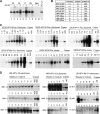
FIG. 1.
FIG. 1.
Southern blot analysis of the transient DNA replication of high- and low-risk mucosal HPV and cutaneous HPV genomes in U2OS cells. (A) Analysis of HPV-16 genome replication in U2OS cells. Episomal DNA was extracted at the indicated time points from U2OS cells that had been transfected with 1 μg (lanes 1 to 4), 2 μg (lanes 5 to 8), or 5 μg (lanes 9 to 12) of recircularized HPV-16 genomes. The DNA was then purified and treated with a linearizing enzyme (BamHI) and DpnI to eliminate the input DNA. The samples were loaded onto a 0.8% agarose gel, separated, transferred to nylon membranes, and hybridized with radiolabeled HPV-16 genome-specific probe as described in Materials and Methods. The episomal DNA extracted from the carrier DNA-transfected U2OS cells is in lane 13. The marker (lane 14) for the replication of the linearized 8-kb fragment of the HPV-16 genome is also indicated by an arrowhead. The DpnI-cleaved input HPV-16 DNA is in lane 15. fr., fragmentation, or cleaving. (B) Analysis of the transient DNA replication of recircularized HPV-6b, -11, and -18 genomes. Religated genomic DNAs of HPV-6b, -11, and -18 (5 μg) were introduced into U2OS cells by electroporation. The episomal DNA was isolated and treated as described above with the exception that subtype-specific hybridization probes were used for each virus. Lanes 1 to 4 represent analyses of HPV-6b DNA replication at the indicated time points. Lane 5 is the marker for the replicating unit size of the HPV-6b genome. Lanes 6 to 9 represent the analyses of HPV-11 genome replication. Lane 10 represents the linear marker for HPV-11 replication. Lanes 11 to 14 represent the analyses of HPV-18 genome replication. Lane 15 is the linear marker for the HPV-18 replicating genome. Lane 16 represents the EcoRI- and DpnI-cleaved input HPV-18 DNA. (C) Transient replication of the HPV-5 genome in U2OS cells. Shown is analysis of replication by restriction with the linearizing enzyme SacI. Lanes 1 to 4, 5 to 8, and 9 to 12 represent cells transfected with 2 μg, 5 μg, or 10 μg, respectively, of recircularized HPV-5 genomes as described in Materials and Methods. Lane 13 shows the analysis of the carrier-transfected cells. Lane 14 is the linear marker for HPV-5 replication. Lane 15 is the SacI- and DpnI-cleaved marker for the input HPV-5 DNA. (D) Transient replication of the HPV-8 genome in U2OS cells. Analysis of replication by restriction with the linearizing enzyme BamHI. Lanes 1 to 4, 5 to 8, and 9 to 12 represent cells transfected with 2 μg, 5 μg, or 10 μg, respectively, of recircularized HPV-8 genomes. Lane 13 shows the analysis of the carrier-transfected cells. Lane 14 shows the linear marker for HPV-8 replication. Lane 15 is the BamHI- and DpnI-cleaved marker for the input HPV-8 DNA. (E) Mutants in two of the early open reading frames, ORFs for E1 and E2, are deficient for transient replication for HPV-18 genomes in U2OS cells. Two micrograms of the viral genome carrying mutations in the E1 or E2 ORF (described in Materials and Methods; a scheme is shown in Fig. 3H, with the names of plasmids shortened) was tested in transient-replication assays separately, in combination with each other, or in complementation with expression vectors for E1 or E2. The mutants alone failed to replicate (lanes 5 and 6, 9 and 10, 15 and 16, and 21 and 22). Replication competence can be restored to E1 and E2 mutant genomes through complementation with each other (lanes 11 and 12, 17 and 18, and 23 and 24) or by addition of appropriate expression vectors (lanes 7 and 8 for the E2 mutant and 13 and 14, 19 and 20, and 25 and 26 for E1 mutants).
FIG. 2.
FIG. 2.
Establishment and maintenance of stable replication of HPV genomes in U2OS cells. HPV-6b, -11, -16, -18, -5, and -8 recircularized genomes (5 μg), carrier plasmid (AraD; 5 μg), and Eco0109I-linearized pNeo-EGFP or EcoRI-linearized pBabeNeo (2 μg) were cotransfected into U2OS cells by electroporation as described in Materials and Methods. Single-cell colonies were picked, expanded, and analyzed using Southern blotting. (A) Analysis of the episomal DNA extracted from HPV-18-, -16-, -11-, and -6b-transfected U2OS cells after 3 weeks of cultivation in the absence (−) or presence (+) of G418. The extracted DNA was purified and cleaved with EcoRI and DpnI for HPV-18 (lanes 1 and 2) and with BamHI and DpnI for HPV-16, -11, and -6b (lanes 3 to 8). DNA from cells transfected with pNeo-EGFP as negative control lanes are also shown (Neo, lanes 9 and 10). M. Lin., position of linear 8-kb HPV fragment. Mixed radiolabeled HPV genomes were used for probes. (B) Stable transfectants and isolated HPV-positive subclones. Two types of linearized selection markers were used, pNeo-EGFP and pBabeNeo (Neo). The different HPV-positive cell lines were isolated from each of the HPV transfection series, the total number of HPV-positive subclones and the yields of HPV-positive subclones are indicated for each HPV type. Collections of these cell lines have been generated. (C) Screening of stable transfectants by Southern blot analysis. Series of identified HPV-positive (Pos.) cell lines for HR-HPV-18 and -16, LR-HPV-11 and -6b, and skin papillomavirus HPV-8 and -5 genomes are grouped and marked above the blocks. Total DNA (10 μg) was used for Southern blotting with appropriate HPV-specific probes. DNA was linearized with EcoRI for the HPV-18 transfection series; with BamHI for the HPV-16, -11, -6b, and -8 transfection series; and with SacI for HPV-5. Subclone numbers for the appropriate HPV types are indicated above the lanes in every series and markers. Calculated viral genome copy numbers per haploid host genome are indicated on the right. Variability in the HPV copy numbers was noticeable in every group. (D) Stability assay for the HR- and LR-HPV subgenomes. Selected cell lines were cultivated for an additional 6 to 11 weeks (indicated by the numbers above the lanes) under regular monolayer culture conditions after the cells were determined to be HPV positive. Total-DNA samples were collected after every week of cultivation and loaded in equal amounts (2 μg for high-copy-number HPV plasmids and 10 μg for low-copy-number HPVs). The linearized total DNA (enzymes are indicated in the previous figure) was analyzed by Southern blotting using appropriate HPV-specific probes. Examples of stable HPV-18-positive cell lines are shown: HPV-18 subclones 18#1.13 and 18#1.4, HPV-16 subclones 16#2.5 and 16#3.16, and HPV-6b subclone 6b#11. Most of the other tested HR-HPV subclones possessed the same type of stability during the tested time period. Examples of clear off for HPV DNA (unstable) were more characteristic in the case of LR-HPVs, as demonstrated for HPV-6b subclone 6b#41, but HPV DNA was also lost from one HPV-16 subclone, 16#3.3. Subclones with stable nonspecific bands (such as 18#1.10), indicating early integration events, were rare. Integration, which is not characteristic of LR-HPVs, was detected for one LR-HPV subclone at later time points (11#3.13). Skin-related subclones are being examined further.
FIG. 3.
FIG. 3.
Induction of DNA amplification in HPV-positive cell lines maintained under dense culture conditions. (A to E) The HPV-18-positive cell line 18#1.13 was grown in a regular monolayer until the cells were equally distributed (1 × 10⁶ cells were seeded onto each of six 100-mm culture dishes) for additional cultivation under dense culture conditions. The medium (IMDM) was changed after every second day (or 2 ml of fresh culture medium was added after every 2 days). The time points for analyses were taken the day after the medium was changed or added during the 12-day growth period, with 2-day intervals. (A) Growth curves of untransfected U2OS cells and the HPV-18-positive cell line 18#1.13. The time-dependent growth of the cells during establishment of the dense cell cultures was analyzed at 2-day intervals for 12 days. The cells were counted with an Invitrogen Countess cell counter. (B) Amounts of total DNA in the time series. Total DNA was isolated by standard procedures, and DNA concentrations were measured using an ND-1000 NanoDrop spectrophotometer. The amounts of total DNA isolated per sample collected every 4 days during one of the 12-day growth periods from U2OS cells and 18#1.13 cells are shown in the bar graph. (C) Induction of DNA amplification under dense culture conditions demonstrated by Southern blot analysis of the HPV-18-positive cell line 18#1.13. Total cellular DNA was isolated at 2-day intervals during the 12-day growth period and analyzed. Equal amounts of DNA (2 μg) were digested with EcoRI and analyzed by Southern blotting with an HPV-18 genome-specific probe. The induction of DNA amplification was demonstrated by the increase of the replication signal over time. A standard series of marker lanes to calculate the copy number per haploid host genome is on the right. (D) Relative HPV-18 DNA copy numbers during amplification. The intensities of the amplification signals from two assays of the 18#1.13 cell line grown in dense cultures were measured using a PhosphorImager and ImageQuant software. The HPV-18 genome copy number was estimated by the standard curve of the marker lanes. The values of two different series were averaged, and the standard deviations are shown by error bars. (E) Increase of the cytokeratin 10 level in confluent HPV-containing U2OS cells. One million of the untreated and HPV18-positive U2OS cells (subclone 18#1.13) were plated on a 10-cm petri dish. The cells were grown with regular feeding of the culture and lysed 1, 4, 8, and 12 days later using Laemmli loading buffer, and the cytokeratin 10 level was measured using Western blot analysis. The lysate from 100,000 cells for every time point was loaded onto an SDS-PAGE gel in sample buffer containing 8 M urea. After separation, the proteins were transferred to a polyvinylidene difluoride (PVDF) membrane, blocked with nonfat milk, and incubated with primary antibody (Abcam) against cytokeratin 10, followed by incubation with anti-mouse horseradish peroxidase (HRP) secondary antibody (LabAs Ltd., Estonia). The cytokeratin 10 level for U2OS cells remained constant over these time points, as for the HPV-18-positive #1.13 cell line a clear increase in the cytokeratin 10 expression levels was detected at the 8- and 12-day time points. (F) RT-PCR analysis of viral mRNA levels at different time points during the induction of amplification of the 18#1.13 cell line. HPV transcript-specific signals were compared to the signal for β-actin. The signals were normalized to time point zero. RT-PCR analysis revealed the upregulation of E1, E2, E6, E7, and L1 mRNA levels. Shown is one of three independent experiments. (G) Examples of the DNA amplification in a selected set of different HPV subclones grown under dense culture conditions. Equally distributed cell samples were analyzed during the 12-day growth period. Southern blot analysis with equally loaded total-DNA samples for HPV-16 subclone 16#2.5, HPV-5 subclone 5#23, HPV-11 subclone 11#74, and HPV-6b subclone 6b#82 are shown. Specific marker standard lanes with calculated HPV copy numbers for each HPV type are on the right. (H) Schematic representation of the locations of sites used for generating E1 and E2 mutants and the locations of RT-PCR primers. Line 1 represents the locations of OliI and StuI restriction sites that were used for the insertion of an oligonucleotide for the generation of mutant genomes. A BcuI site was used to create a frameshift in the E1 ORF by filling the generated overhangs. Line 2 represents the locations of the RT-PCR primers designed to measure the mRNA levels in cells.
FIG. 4.
FIG. 4.
Induction of HPV amplification in U2OS cells detected by FISH. Samples of HPV-18 subclone 18#1.13 were collected on days 2 and 14 after seeding during the amplification assay and were analyzed by FISH (Invitrogen Corporation; TSA Kit 22) as described in Materials and Methods. (A) Uninduced 18#1.13 interphase cells with a weak HPV-18 signal detected on day 2 after seeding. (B) Induction of amplification in 18#1.13 interphase cells with extensive, widespread HPV-18 signals detected 2 weeks after seeding.
FIG. 5.
FIG. 5.
Southern blot analysis for determining the structure of DNA replication intermediates. Abbreviations: mw, ladder of linear fragments; sc, ladder of supercoiled molecules; mc, multimeric circles; L, linear molecules. (A) Detection of HPV-18 DNA species during the first amplification. U2OS cells were cotransfected with 2 μg of religated HPV-18 DNA and carrier DNA. The uncut DNA from Hirt lysates (lanes 1 to 3) or 2 μg of total DNA (lanes 4 to 7) extracted 72, 96, and 168 h and 3 weeks (3w) posttransfection were separated on a 1D agarose gel and analyzed by Southern blotting using an HPV-18 genomic probe. A 7.1-kb circular (circ) HPV-18 E1 expression construct and an 11-kb circular pBR-HPV-18 DNA were used as plasmid DNA markers. (B) DNA status of HPV-18-positive subclone 18#1.13 during the second amplification. Two micrograms of HindIII (noncutter)-treated total DNA from HPV-18-positive cell line 18#1.13 was loaded, and replication intermediates were investigated during the 12-day incubation period in the amplification assay. The 7.1-kb plasmid DNA (HPV-18 E1 expression vector) and 11-kb pUC/HPV-18 plasmid DNA were used as molecular markers. (C) Head-to-tail oligomeric forms of the HPV DNA from HPV-18-positive subclone 18#1.13. The Hirt supernatant and pellet were extracted from cells cultivated under regular growth conditions allowing the stable maintenance of viral genomic material and were subjected to 1D analysis. The Hirt supernatant (whole DNA from a 10-cm plate per lane) and pellet samples (5 μg per lane) were digested with two noncutter enzymes for HPV-18 (HindIII and BglII) (lanes 1 and 2 and 6 and 7) and with three linearizing (lin) single-cutter enzymes for HPV-18 (EcoRI, XmaJI, and BglI) (lanes 3 to 5 and 8 to 10). Samples were analyzed by Southern blotting using an 8-kb linear HPV-18 genomic probe. Size markers for the linear DNA are on the right. All three linearizing enzymes gave the same 8-kb band, indicating that the observed DNA forms had been organized into units by the head-to-tail tandems. (D) The amplification signal comes from extrachromosomal oligomeric forms of HPV DNA. The Hirt supernatant and pellet were prepared from the HPV-18-positive subclone 18#1.13 cultivated under conditions inducing amplification of the viral genomic material (see Materials and Methods). The Hirt supernatant (DNA from 1/5 of the 10-cm plate per lane) and pellet samples (2 μg DNA per lane) were digested with two noncutter enzymes for HPV-18 (HindIII and BglII) (lanes 1 and 2 and 6 and 7) and with three linearizing single-cutter enzymes (EcoRI, XmaJI, and BglI) (lanes 3 to 5 and 8 to 10). Samples were analyzed by Southern blotting using an 8-kb linear HPV-18 genomic probe. Size markers for linear DNA are on the left. Most of the generating HPV DNA structures are in extrachromosomal form or in monomeric and most likely in head-to-tail multimeric forms during passaging of the HPV-positive cells under dense culture conditions. (E) Schematic presentation of the migration of undigested DNA. L, linear molecules; CCC, covalently closed circles; and OC, open circles. The numbers refer to the multimeric state (1, monomers; 2, dimers; etc.). CCC/OC are DNA molecules that migrate as covalently closed circles in the first dimension and that were subsequently nicked and migrated as relaxed circles in the second dimension. catA represents concatemers formed by relaxed circles that are linked in a noncovalent manner. (F to H) Neutral/neutral two-dimensional gel analysis of structures of DNA replication products. The episomal DNA extracted by Hirt lysis from 18#1.13 cells grown as regular (F) or dense (G and H) monolayer cultures is shown. The undigested DNA was separated by 2D electrophoresis (see Materials and Methods) and analyzed by Southern blotting using an HPV-18 genome-specific probe. The size markers of supercoiled DNA are shown in both directions (sc). The presence of an 8-kb circular plasmid is shown by the arrowhead. The generation of additional high-molecular-weight plasmid multimers was also detected during amplification (G and H). The samples shown in panel H were allowed to run for a longer time in the second dimension than the samples shown in panel G for better separation of the molecules in this dimension. (I) Schematic presentation of the migration of replication forks. 1N is the starting spot of the position. 2N is the position at which the 1N molecules that were almost completely replicated would be expected to migrate. (J and K) 2D Southern blots for restriction analysis. LMW DNA from 18#1.13 cells in dense cultures was digested with NcoI and Bpu1102I and analyzed using 2D electrophoresis. An NcoI-Bpu1102I-digested HPV-18 genomic DNA fragment (J) or an XmaJI-AatII-digested HPV-18 genomic DNA fragment (K) was used as a hybridization probe for the 2D Southern blots. Marker lines for the 1-kb ladder (Fermentas) are indicated in both directions. (L) Schematic representation of the circular dimer form of the HPV-18 genome. The initiation of DNA replication occurs at the active ori element and terminates approximately 180° opposite, where the inactive origin is located. The proximal locations of the restriction sites of the enzymes used in the study are also indicated.

Similar articles

Cited by

References

    1. Aaltonen, L. M., T. Wahlstrom, H. Rihkanen, and A. Vaheri. 1998. A novel method to culture laryngeal human papillomavirus-positive epithelial cells produces papilloma-type cytology on collagen rafts. Eur. J. Cancer 34:1111-1116. - PubMed
    1. Adachi, A., T. Kiyono, Y. Hayashi, M. Ohashi, and M. Ishibashi. 1996. Detection of human papillomavirus (HPV) type 47 DNA in malignant lesions from epidermodysplasia verruciformis by protocols for precise typing of related HPV DNAs. J. Clin. Microbiol. 34:369-375. - PMC - PubMed
    1. Andrei, G., S. Duraffour, J. Van den Oord, and R. Snoeck. 2010. Epithelial raft cultures for investigations of virus growth, pathogenesis and efficacy of antiviral agents. Antiviral Res. 85:431-449. - PubMed
    1. Chiang, C. M., G. Dong, T. R. Broker, and L. T. Chow. 1992. Control of human papillomavirus type 11 origin of replication by the E2 family of transcription regulatory proteins. J. Virol. 66:5224-5231. - PMC - PubMed
    1. Chiang, C. M., et al. 1992. Viral E1 and E2 proteins support replication of homologous and heterologous papillomaviral origins. Proc. Natl. Acad. Sci. U. S. A. 89:5799-5803. - PMC - PubMed

Publication types

LinkOut - more resources