. 2011 Apr;85(7):3077-85.
doi: 10.1128/JVI.02257-10. Epub 2011 Jan 19.

Prion propagation in cells expressing PrP glycosylation mutants

Affiliations

Prion propagation in cells expressing PrP glycosylation mutants

Muhammad K Salamat et al. J Virol. 2011 Apr.

Abstract

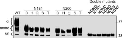
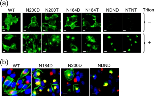
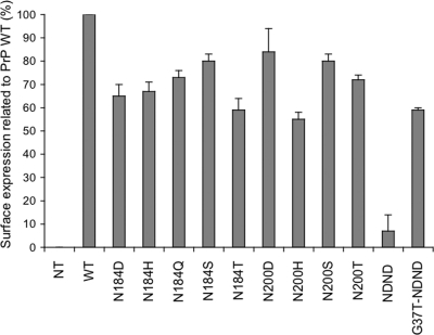
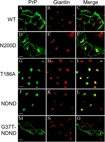
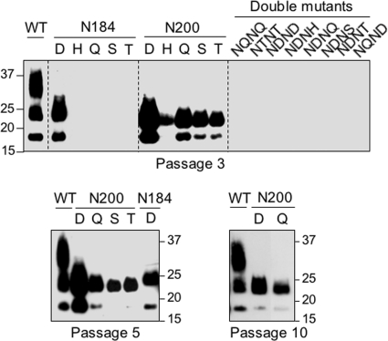
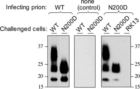
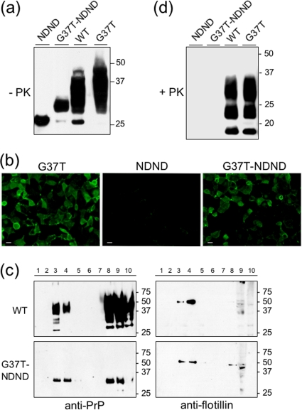
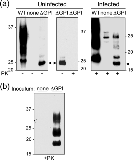
Infection by prions involves conversion of a host-encoded cell surface protein (PrP(C)) to a disease-related isoform (PrP(Sc)). PrP(C) carries two glycosylation sites variably occupied by complex N-glycans, which have been suggested by previous studies to influence the susceptibility to these diseases and to determine characteristics of prion strains. We used the Rov cell system, which is susceptible to sheep prions, to generate a series of PrP(C) glycosylation mutants with mutations at one or both attachment sites. We examined their subcellular trafficking and ability to convert into PrP(Sc) and to sustain stable prion propagation in the absence of wild-type PrP. The susceptibility to infection of mutants monoglycosylated at either site differed dramatically depending on the amino acid substitution. Aglycosylated double mutants showed overaccumulation in the Golgi compartment and failed to be infected. Introduction of an ectopic glycosylation site near the N terminus fully restored cell surface expression of PrP but not convertibility into PrP(Sc), while PrP(C) with three glycosylation sites conferred cell permissiveness to infection similarly to the wild type. In contrast, predominantly aglycosylated molecules with nonmutated N-glycosylation sequons, produced in cells expressing glycosylphosphatidylinositol-anchorless PrP(C), were able to form infectious PrP(Sc). Together our findings suggest that glycosylation is important for efficient trafficking of anchored PrP to the cell surface and sustained prion propagation. However, properly trafficked glycosylation mutants were not necessarily prone to conversion, thus making it difficult in such studies to discern whether the amino acid changes or glycan chain removal most influences the permissiveness to prion infection.


Figures

FIG. 1.
FIG. 1.
Expression of PrP glycosylation mutants in Rov cell cultures. Immunoblotting analysis with 4F2 antibody of Rov cells that expressed PrP mutants with asparagine substitutions at codon 184 or 200 (sheep sequence numbering) or at both codons (double mutants) is shown. The positions of the diglycosylated, monoglycosylated, and unglycosylated forms of PrP are indicated on the left. While diglycosylated species predominate with wild-type (WT) PrP, monoglycosylated species become the major component with single mutants. Double mutants show only one band corresponding to unglycosylated PrP. Molecular weights (in thousands) are indicated on the right.
FIG. 2.
FIG. 2.
Subcellular localization of PrP glycosylation mutants. (a) Immunofluorescence staining of Triton X-100-permeabilized and nonpermeabilized cells using 4F2 antibody. Cells expressing monoglycosylated PrP mutants displayed a typical cell surface labeling together with a regionalized intracellular signal, whereas in cells expressing double PrP mutants the labeling was essentially intracellular. (b) Double immunofluorescence staining of PrP (in green) and of a marker of the Golgi apparatus (giantin, in red). Bars, 10 μm.
FIG. 3.
FIG. 3.
Comparative cell surface expression of PrP glycosylation mutants as determined by flow cytometry. The expression levels of the mutants are provided as percentage relative to wild-type PrP (means and standard deviations from three experiments; 4F2 antibody). NT, nontransfected RK13 cells. G37T-NDND designates a double mutant with an extra glycosylation site, which is described in the text.
FIG. 4.
FIG. 4.
Accumulation of PrPC mutants in the Golgi compartment. PrP and giantin were labeled as for Fig. 2, and fluorescence emission was examined by confocal microscopy. Z-axis planes are shown. The mutants analyzed (D to O) are indicated on the left (for G37T-NDND, see the Fig. 3 legend). Bars, 10 μM.
FIG. 5.
FIG. 5.
Prion infection of Rov cells expressing PrP glycosylation mutants. Cultures exposed to an infectious inoculum of the 127S ovine prion strain were tested for the presence of PK-resistant PrP by immunoblotting using Sha31 antibody. Results of a typical experiment are shown, and the number of passages postexposure is indicated. The gels were loaded with 250 μg or 25 μg of PK-digested proteins per lane for the mutant or wild-type PrP-containing samples, respectively.
FIG. 6.
FIG. 6.
Mutant PrP-expressing cells can propagate prion infectivity. Cellular homogenates prepared from N200D mutant or wild-type PrP Rov cell cultures at six passages postexposure were used to infect new recipient cell cultures, as indicated. These cultures were assayed for the presence of PK-resistant PrP at three passages postexposure.
FIG. 7.
FIG. 7.
Expression and permissiveness to prion infection of PrP mutants with an extra glycosylation site. (a) Immunoblot showing the PrPC glycoform profiles of mutants bearing either three glycosylation sites (G37T) or the extra glycosylation site only (G37T-NDND). The artificial N-linked glycosylation sequon created by replacement of a glycine by a threonine at position 37 was truly functional, as evidenced by the modification of the glycoform profiles of G37T-NDND and of G37T mutants compared to NDND and wild-type PrP, respectively. (b) Expression of G37T-NDND PrP at the cell surface. Immunofluorescence on living cells using 4F2 antibody is shown. G37T and G37T-NDND PrPs show similar cell surface labeling, while the NDND mutant is essentially negative. Bars, 20 μM. (c) G37T-NDND mutant and wild-type PrPs are both associated with lipid rafts. An immunoblot analysis performed on step sucrose gradient fractions using anti-PrP and antiflotillin antibodies is shown. Fractions 3 and 4 correspond to the interface of 5% and 30% sucrose cushions. (d) Permissiveness to de novo prion infection of the G37T PrP mutants. An immunoblot with PK-treated cell lysates from cultures at passage 3 postexposure is shown (Sha31 antibody). No PrPres signal is visible in G37T-NDND mutant-expressing cells, although G37T mutant and wild-type PrP-expressing cells accumulate PrPres at similar levels.
FIG. 8.
FIG. 8.
Ability of anchorless PrP to form PrPSc. (a) Left panel, immunoblot analysis of Rov cell lysates expressing ΔGPI or wild-type PrPC; middle panel, PK sensitivity of ΔGPI PrP expressed in mock-infected cells; right panel, PK resistance of ΔGPI PrP in cultures exposed to infectious prion inoculum (see Materials and Methods; Sha31 antibody). None, nontransfected, parental RK13 cells. Note the faster migration of ΔGPI (arrowheads) compared to wild-type PrP. Upper bands in the infected, PK-treated nontransfected, and ΔGPI samples represent nonspecific background. (b) ΔGPI PrPSc is infectious. Cellular material prepared from lysates of infected cultures such as shown in panel a (either RK13 [none] or ΔGPI cells) was inoculated into cells expressing wild-type PrPC. PK-treated samples from infected cells at the second passage are shown.
FIG. 9.
FIG. 9.
Permissiveness to prion infection of PrP mutants with an altered glycoform pattern. Human PrP mutations reported to alter the PrPC glycosylation pattern were introduced into the sheep sequence, as indicated (ovine PrP numbering). Immunoblot analysis for PrPC (a) (mock-infected culture, 4F2 antibody) and PrPres (b) (prion-infected cultures at third passage postexposure, Sha31 antibody) is shown. Note that the F210V mutant but not the F210S mutant conferred susceptibility to infection.

Similar articles

Cited by

References

    1. Aguzzi, A., F. Baumann, and J. Bremer. 2008. The prion's elusive reason for being. Annu. Rev. Neurosci. 31:439-477. - PubMed
    1. Ashok, A., and R. S. Hegde. 2009. Selective processing and metabolism of disease-causing mutant prion proteins. PLoS Pathog. 5:e1000479. - PMC - PubMed
    1. Beringue, V., et al. 2003. Regional heterogeneity of cellular prion protein isoforms in the mouse brain. Brain 126:2065-2073. - PubMed
    1. Beringue, V., J. L. Vilotte, and H. Laude. 2008. Prion agent diversity and species barrier. Vet. Res. 39:47. - PubMed
    1. Birkett, C. R., et al. 2001. Scrapie strains maintain biological phenotypes on propagation in a cell line in culture. EMBO J. 20:3351-3358. - PMC - PubMed

Publication types