. 2011 Apr;85(7):3270-82.
doi: 10.1128/JVI.02243-10. Epub 2011 Jan 19.

M062 is a host range factor essential for myxoma virus pathogenesis and functions as an antagonist of host SAMD9 in human cells

Affiliations

M062 is a host range factor essential for myxoma virus pathogenesis and functions as an antagonist of host SAMD9 in human cells

Jia Liu et al. J Virol. 2011 Apr.

Abstract

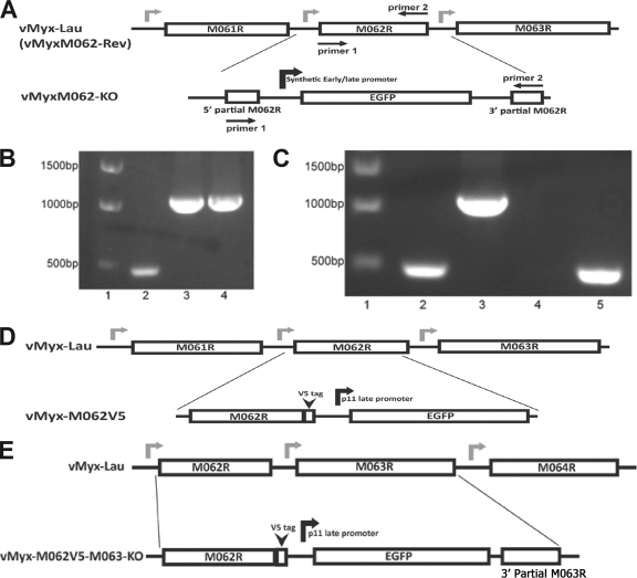
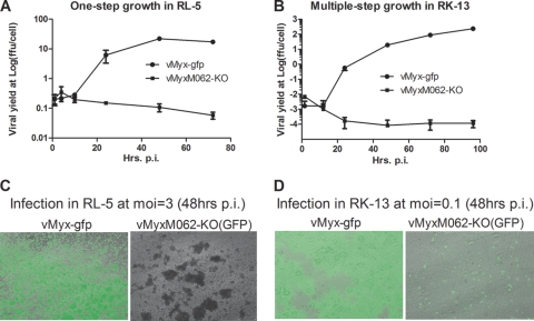
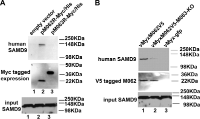
Myxoma virus (MYXV) M062R is a functional homolog of the C7L family of host range genes from orthopoxviruses. We constructed a targeted M062R-knockout-MYXV (vMyxM062-KO) and characterized its properties in vitro and in vivo. In European rabbits, infection by vMyxM062-KO was completely asymptomatic. The surviving rabbits did not gain full protection against the subsequent lethal-dose challenge with wild-type MYXV. We also looked for cellular tropism defects in a variety of cultured cells. In all of the rabbit cells tested, vMyxM062-KO conducts an abortive infection, although it initiates viral DNA replication. In many, but not all, human cancer cells that are permissive for wild-type MYXV, vMyxM062-KO exhibited a profound replication defect. We categorized human cells tested into two groups: (i) type A, which support productive replication for wild-type MYXV but are unable to produce significant levels of progeny virus by vMyxM062-KO, and (ii) type B, which are permissive to infections by both wild-type MYXV and vMyxM062-KO. Furthermore, using proteomic strategies, we identified sterile α motif domain containing 9 (SAMD9), an interferon-regulated cellular protein implicated in human inflammatory disorders, as a unique host binding partner of M062 in human cells. Significantly, knocking down SAMD9 in type A human cancer cells led to a substantial rescue of vMyxM062-KO infection. In summary, M062 is a novel host range factor that controls productive MYXV replication in rabbit cells and in a wide variety of human cells. M062 also binds and antagonizes cellular SAMD9 in human cells, suggesting that SAMD9 is a novel innate antiviral factor against poxviruses.


Figures

FIG. 1.
FIG. 1.
Construction and confirmations of recombinant MYXVs. (A) Construction of vMyxM062-KO. The central 78% of MYXV M062R sequence was replaced by an eGFP expression cassette under a viral early/late promoter using the three-element recombination of the MultiSite Gateway system. The revertant virus of vMyxM062-KO was constructed by replacing the eGFP expression cassette in the knockout virus with an intact M062R sequence. (B) Confirmation of purified vMyxM062-KO. PCR analysis using primers 1 and 2 (Table 3) designed to recognize M062R coding sequences (shown in panel A) will generate a 474-bp fragment with wild-type MYXV viral DNA as a template (lane 2) and a 1,077-bp fragment using either the recombination plasmid (lane 3) or purified vMyxM062-KO viral DNA (lane 4) as a template. The molecular markers are in lane 1. (C) Confirmation of the purified vMyxM062-Rev. The set of screening primers used in panel B was used to determine whether vMyxM062-Rev contained any contaminating vMyxM062-KO. Lane 1, molecular marker; lane 2, vMyx-Lau viral DNA; 3, vMyxM062-KO DNA; 4, no template DNA; 5, DNA of vMyxM062-Rev. (D) Construction of vMyx-M062V5. Using the MultiSite Gateway system, a V5 epitope sequence was inserted just before the stop codon of the M062R. In addition, a cassette containing a VACV p11 late promoter-driven eGFP expression was inserted following the V5-tagged M062R and in front of the intact M063R promoter region. Primers 1 and 3 (Table 3) were used for screening during the purification process. (E) The construction of vMyx-M062V5-M063-KO. MultiSite Gateway system was used to insert the V5 sequence before the stop codon of M062R, VACV p11 late promoter-driven eGFP, and 115 bp of the 3′-terminal sequence of M063R in front of an intact M064R promoter sequence. Primers 1 and 3 were used for screening during the process of purification.
FIG. 2.
FIG. 2.
Infection by vMyxM062-KO in rabbit cells is abortive. (A) One-step growth curve of MYXV infection in RL-5 rabbit T cells. The rabbit CD4+ T cell line RL-5 was infected with either vMyx-gfp or vMyxM062-KO at an MOI of 3, and total cell lysates were collected at given time points (1, 4, 10, 24, 48, and 72 h p.i.). The viral yield at each time point was estimated by titrating on BSC-40 cells, and titrations were performed in triplicate to calculate the average titer at each time point, while an error bar represents the ± the standard deviation (SD). Shown is a representative one-step growth curve of three independent experiments. (B) Multiple-step growth of MYXV in RK-13 rabbit kidney cells. Either vMyx-gfp or vMyxM062-KO was used to infect RK-13 at an MOI of 0.1, at given time points (1.5, 12, 24, 48, 72, and 96 h p.i.) total cell lysates were harvested for titration as described in panel A. Shown is a representative multiple-step growth curve from three independent experiments. (C) Fluorescence microscope images of infected RL-5 cells. Images of infected cells from panel A were taken using an inverted fluorescence microscope (Leica DMI6000B) using a lens with ×10 magnification at 48 h p.i. The pictures shown are merged images from both GFP and bright fields. Left side, infection by vMyx-gfp; right side, infection by vMyxM062-KO. (D) Fluorescent images of infected RK-13 cells. At 48 h p.i., pictures of infected cells from panel B were taken under both GFP filter and bright field. Merged images are shown. Left side, infection by vMyx-gfp; right side, infection by vMyxM062-KO.
FIG. 3.
FIG. 3.
M062 is an early/late gene product and is essential for MYXV infection in rabbit cells. (A) Viral DNA is detected in RK-13 cells infected with vMyxM062-KO. RK-13 cells were infected with either vMyx-gfp or vMyxM062-KO at an MOI of 5, and cells were harvested at 2, 4, 8, 12, and 24 h p.i. Total DNA was extracted and real-time PCR was conducted using Sybr green real-time PCR system. Primer sets of MYXV M071L and the 18S ribosomal genes from O. cuniculus were designed to selectively amplify MYXV DNA and rabbit cellular DNA, respectively. Real-time PCR for M071L or 18S at each time point for each infection was conducted in triplicate. The average CT from the triplicate was used for the calculation of the ratio comparison. The comparative CT method or the 2−ΔΔCT method was used to calculate the relative fold increase (RFI) of viral DNA level during infection by vMyx-gfp or vMyxM062-KO. For each infection, the fold increase of sample (viral) DNA level at each time point was measured against the sample DNA level at 2 h p.i., which was set artificially as 1-fold. Curve shown is a summary of results from two independent experiments, and error bars represent ± the SD. (B) Infection of RK-13 cells with vMyxM062-KO led to an abortive infection with no late viral gene expression. RK-13 cells were mock treated (lane 1) or infected with either vMyx-gfp or vMyxM062-KO at an MOI of 5 in the absence or the presence of AraC. At 4, 8, 12, or 24 h p.i., monolayer cells were lysed to measure total protein concentration. Total protein (50 μg) from each sample was separated using 12% SDS-PAGE, transferred to a PVDF membrane, and analyzed by Western blotting. The membrane was first probed for an antibody raised against a late protein of MYXV (SERP-1), stripped, and probed for β-actin as a loading control. Lanes 1 and 11, molecular marker; lane 2, mock infection without AraC; lanes 3, 4, and 5, infection by vMyxM062-KO in the absence of AraC harvested at 4, 8, and 12 h p.i., respectively; lanes 6, 7, and 8, infection by vMyx-gfp in the absence of AraC harvested at 4, 8, 12 h p.i., respectively; lanes 9 and 10, infection by vMyx-gfp in the presence of AraC harvested at 8 and 12 h p.i., respectively; lanes 12 and 13, infection by vMyx-gfp and vMyxM062-KO, respectively, harvested at 24 h in the absence of AraC. (C) M062 mRNA is no longer expressed after vMyxM062-KO infection in rabbit cells. RK-13 cells were infected with either vMyx-gfp or vMyxM062-KO at an MOI of 5. At 1, 2, 4, 8, and 12 h p.i. for vMyx-gfp infection or at 2, 8, and 12 h p.i. for vMyxM062-KO infection or a mock infection sample (as controls for “no M062 cDNA”), cells were harvested to extract total RNA, and reverse transcription was conducted to produce cDNA. Sybr green real-time PCR was conducted to measure the M062 cDNA level using a primer set shown in Table 3, and O. cuniculus 18S cDNA was also measured as an internal control. Comparative CT method was used to calculate the level increase of M062 mRNA during vMyx-gfp infection, while no stable M062 mRNA can be detected at any time point after infection by vMyxM062-KO or in the mock-treated sample. (D) M062 protein is expressed as an early/late gene product during wild-type MYXV infection in rabbit cells. RK-13 cells were infected with vMyxM062V5 or mock infected at an MOI of 5 in the presence or absence of AraC. At 0, 1, 2, 4, 8, 12, and 24 h p.i. in the absence of AraC and at 8, 12, and 24 h p.i. in the presence of AraC, cells were harvested for analysis by Western blot, and 50 μg of total protein from each sample was separated by a 12% SDS-PAGE, transferred to a PVDF membrane, and analyzed by Western blotting with a V5 antibody. The blot was then stripped and probed for M063 (an early/late MYXV protein), M-T7 (an early/late viral protein), and SERP-1 (a late viral protein), respectively, and finally β-actin as loading control. Lanes 1 to 7, infection at 0, 1, 2, 4, 8, 12, and 24 h p.i., respectively, in the absence of AraC; lanes 8, 9, and 10, infection at 8, 12, and 24 h p.i., respectively, in the presence of AraC.
FIG. 4.
FIG. 4.
M062 is essential for MYXV pathogenesis in European rabbits. (A) Comparison of in vivo pathogenesis showed that vMyxM062-KO can no longer cause productive infection in rabbits. A total of 1,000 FFU of vMyx-Lau (n = 5), vMyxM062-KO (n = 4), or vMyxM062-Rev (n = 3) were diluted in 100 μl of PBS and inoculated intradermally into the left flank of European rabbits. Another group of rabbits (n = 3) were inoculated with PBS only. The overall physical condition and disease progression were evaluated daily in a clinical score system, and the average daily score was calculated. The error bar in the chart represents ± the SD within each group. (B) MYXV without M062 can no longer cause lethal myxomatosis. The results of the in vivo study described in panel A were analyzed for survival rates among the groups. The daily percentage of survival in each group was plotted to generate the survival curve.
FIG. 5.
FIG. 5.
M062 is essential for MYXV infection in many diverse human cells. (A) Multiple-step growth curves of MYXV in U87 glioma cells. U87 cells were infected at an MOI of 0.1 with either vMyx-gfp or vMyxM062-KO. At given time points (12, 24, 48, and 72 h p.i. for both viruses and up to 96 h p.i. for vMyxM062-KO), total cell lysates were harvested for titration. Error bars represent ± the SD generated from titration in triplicate, and the results of a representative growth curve of two independent experiments are shown. (B) One-step growth curves of MYXV infection in U87 cells. U87 cells were infected at an MOI of 3 with vMyx-gfp or vMyxM062-KO. Cell lysates were harvested at 2, 4, 12, 24, and 57 h p.i. for titration, and error bars represent ± the SD generated from titration in triplicate. The results of a representative growth curve from two independent experiments are shown. (C) Multiple-step growth curves of MYXV in Huh7 cells. Experiments were carried out as described for panel A. (D) One-step growth curves of MYXV in Huh7. Experiments were carried out as described for panel B. (E) Multiple-step growth curves in HepG2 cells. Experiments were carried out as described for panel A. (F) One-step growth curves in HepG2 cells. Experiments were carried out as described for panel B.
FIG. 6.
FIG. 6.
M062 cannot be fully compensated for by other functional homologs of C7L family, and M062 has a unique interaction to a human host factor, SAMD9. (A) Transient expression of C7L family members can partially rescue vMyxM062-KO infection in human cells. HEK293 cells were infected with vMyxM062-KO at an MOI of 1 for 1 h, followed by transient transfection of the plasmids pYLDV-67R, pVACV-C7L, and pMYXV-M062R, respectively, or were mock transfected. The construction of these plasmids has been previously described (16). In these plasmids, the sequence of the VACV-C7L native promoter drives the expression of V5 tagged YLDV-67R, VACV-C7L, or MYXV-M062R. At 48 h posttransfection, pictures were taken using a fluorescence microscope with a lens of ×10 magnification under both a GFP filter and a bright field. Merged images are shown. (B) The interaction with human SAMD9 protein is unique and specific to M062 and does not occur with other functional homologs of the C7 family. Cell lysates from the experiment described in panel A were harvested at 72 h posttransfection. Before co-IP using the agarose-conjugated anti-V5 antibody, a 1/50 volume of total protein lysate from each sample was saved to prepare a Western blot to reveal the input human SAMD9 (bottom panel). Proteins associated with V5-tagged proteins were then separated on 10% SDS-PAGE, transferred to a PVDF membrane, and immunoblotted. The blot was first probed for human SAMD9 (top panel), stripped, and then probed for V5-tagged protein expression (middle panel). Lanes 1 to 4 represent cells infected with vMyxM062-KO and treated with mock transfection, transfected with pYLDV-67R, pVACV-C7L, or pMYXV-62R, respectively. (C) Antibody recognizing human SAMD9 for co-IP also confirms the interaction with M062 and M063. Human HCT116 cells were mock infected (lane 1), infected with vMyx-gfp (lane 2), or infected with vMyxM062V5 (lane 3) at an MOI of 1 for 72 h. Before co-IP, 1% volume of each cell lysate was used for Western blotting to show the input SAMD9. Proteins associated with SAMD9 were separated on 10% SDS-PAGE for immunoblotting as previously described for panel B. The blot was first probed for V5-tagged protein, stripped, and probed for M063 and then SAMD9. (D) In RK-13 rabbit cells, the expression of VACV-C7 or YLDV-67 did not compensate for the loss of M062R gene during the infection by vMyxM062-KO. RK-13 cells were infected with vMyxM062-KO at an MOI of 1 for 1 h before they were mock transfected or transfected with pMYXV-M062RV5, pVACV-C7LV5, or pYLDV-67RV5, respectively. At 72 h p.i., the cells were examined by using a fluorescence microscope, and images were taken using a lens of ×10 magnification. Merged green fluorescent and bright-field images are shown in subpanel a. The total protein (50 μg) was separated by 12% SDS-PAGE, transferred to a PVDF membrane, and analyzed by Western blotting. The membrane was probed first using anti-SERP-1 antibody, stripped, and then probed to monitor V5-tagged protein expression (not shown). At last, the membrane was stripped and probed for β-actin expression as a loading control. The results of Western blot analyses are shown in subpanel b. Lane 1, RK-13 cells were infected with vMyxM062-KO and mock transfected; lanes 2 to 4, RK-13 cells were infected with vMyxM062-KO and transfected with pMYXV-M062RV5, pVACV-C7LV5, or pYLDV-67RV5, respectively; lane 5, HeLa cells were infected with vMyxM062-KO at an MOI of 1 for 72 h as a positive control for SERP-1 expression.
FIG. 7.
FIG. 7.
In human cells M062, but not M063, binds to endogenous SAMD9 protein, but the presence of M063 during MYXV infection enhances the interaction of M062 and SAMD9. (A) M062 binds to human SAMD9. HeLa cells were transfected with empty vector (pCDNA3.1-Myc/His) (lane 1), pM062R-Myc/His (lane 2), or pM063R-Myc/His (lane 3) for 48 h. Cells were lysed, and before a pull-down experiment using Ni-NTA beads was constructed, 1/50 volume of the cell lysate was saved for Western blotting to show the input SAMD9 (bottom panel). Proteins associated with the indicated transfected His-tagged proteins were pulled down, washed, and eluted from the beads. The eluted protein was separated by 10% SDS-PAGE and transferred to a PVDF membrane for immunoblotting. The membrane was first probed with anti-human SAMD9 antibody, stripped, and then probed with anti-myc antibody for myc-tagged protein expression. The SAMD9 protein was detected at a molecular mass of ∼148 kDa only in cells transfected with pM062R-Myc/His (top panel). The M063-myc tagged protein was detected at the observed molecular mass of 29.2 kDa, while the M062-myc tagged protein was detected at 23.7 kDa (middle panel). (B) The expression of M063 protein enhances the binding between M062 and SAMD9 in human cells during MYXV infection. U87 cells were infected with vMyxM062V5 (lane 1), vMyxM062V5-M063-KO (lane 2), or vMyx-gfp (lane 3) at an MOI of 1 for 48 h. Cells were harvested and, before co-IP using anti-V5 antibody was performed, a 1/50 volume of total cell lysate was saved for Western blotting to show SAMD9 input (bottom panel). Precipitated proteins were separated by 8% SDS-PAGE and transferred for an immunoblot procedure. The blot was first probed for SAMD9 expression (top panel). After stripping, it was then probed with anti-V5 antibody (middle panel).
FIG. 8.
FIG. 8.
Reducing the expression of endogenous human SAMD9 can rescue vMyxM062-KO infection in nonpermissive cells. (A) Knocking down the expression of endogenous SAMD9 in U87 can rescue late viral gene expression following vMyxM062-KO infection. U87 lines with constitutive SAMD9 knockdown were constructed by infecting parental U87 with a lentivirus packaged with shRNAs targeting SAMD9 mRNA and by selecting for transduced clones with the highest knockdown at the protein level. In this experiment, the number 9 clone, designated U87 SAMD9-shRNA-9, was used. A control U87 cell line, designated U87 control-shRNA, was constructed by infecting U87 cells with a lentivirus packaged with control shRNAs and selecting for stably transduced cells. Parental U87, U87 control shRNA, and U87 SAMD9-shRNA-9 were infected with vMyxM062-KO at an MOI of 5. Cell lysates were harvested at 8, 12, and 24 h p.i., and 25 μg of total protein of each sample was separated by 12% SDS-PAGE and then analyzed by Western blotting. The blot was first probed against SERP-1, and after stripping it was probed against β-actin as a loading control. (B) Cell lines with reduced SAMD9 expression showed rescue of productive infection by vMyxM062-KO. One-step growth of vMyxM062-KO was conducted in parental U87, U87 control-shRNA, U87 SAMD9-shRNA-7, and U87 SAMD9-shRNA-9 at an MOI of 5. At 3, 8, 24, and 48 h p.i., total cell lysates were collected for titration. Error bars represent ± the SD calculated from infections performed in triplicate. (C) Transient knockdown of endogenous SAMD9 improved the M062 knockout MYXV yield in Huh7 cells. Huh7 cells were treated in triplicate as follows: (i) transiently transfected with control siRNA (lane 2) or (ii) siRNAs targeting human SAMD9 (final concentration of 100 nM) (lane 3) or (iii) treated with only transfection reagent (mock) (lane 1) for 48 h before infection with vMyxM062-KO at an MOI of 3 for 48 h. Cell lysates were harvested for titration, and error bars represent ± the SD calculated from viral yields in triplicate in each treatment. One-way analysis of variance was used to analyze the statistical significance among the different viral yields in each group (P = 0.0016) and Tukey's multiple-comparison test was conducted to conclude that viral yield from SAMD9 siRNA-transfected group (labeled with an asterisk [*]) is significantly different from the other two groups. (D) Late gene expression of MYXV after infection increased by knocking down SAMD9 expression in Huh7 cells. Huh7 cells were infected with a lentivirus packaged with either a control shRNA or with SAMD9 shRNAs, followed by puromycin selection to enrich for the transduced cells. Significant knockdown of SAMD9 was observed in cells transduced with SAMD9 shRNA (labeled as “S9”), while the SAMD9 protein level is intact in control shRNA transduced cells (labeled as “C”). Infection by vMyxM062-KO was conducted at an MOI of 5 in both Huh7-S9 (SAMD9 knockdown) and Huh7-C (control shRNA) cells. At 4, 8, 12, and 26 h p.i., cell lysates were collected, along with cell lysate from the mock-infected cells (Huh7-C) (lane 1), and 25 μg of total protein was separated by 12% SDS-PAGE and analyzed by Western blotting. The blot was first probed for SERP-1 (top panel), stripped, and then probed for β-actin (bottom panel).

Similar articles

Cited by

References

    1. Adams, M. M., B. H. van Leeuwen, G. McFadden, and P. J. Kerr. 2008. Construction and testing of a novel host-range defective myxoma virus vaccine with the M063 gene inactivated that is non-permissive for replication in rabbit cells. Vet. Res. 39:60. - PubMed
    1. Backes, S., et al. 2010. Viral host-range factor C7 or K1 is essential for modified vaccinia virus Ankara late gene expression in human and murine cells, irrespective of their capacity to inhibit protein kinase R-mediated phosphorylation of eukaryotic translation initiation factor 2α. J. Gen. Virol. 91:470-482. - PubMed
    1. Barrett, J. W., et al. 2007. Identification of host range mutants of myxoma virus with altered oncolytic potential in human glioma cells. J. Neurovirol. 13:549-560. - PubMed
    1. Barrett, J. W., et al. 2007. Myxoma virus M063R is a host range gene essential for virus replication in rabbit cells. Virology 361:123-132. - PubMed
    1. Barry, M., S. F. Lee, L. Boshkov, and G. McFadden. 1995. Myxoma virus induces extensive CD4 downregulation and dissociation of p56lck in infected rabbit CD4+ T lymphocytes. J. Virol. 69:5243-5251. - PMC - PubMed

Publication types

MeSH terms