. 2011 Apr;85(7):3294-305.
doi: 10.1128/JVI.01994-10. Epub 2011 Jan 19.

Processing and localization of the african swine fever virus CD2v transmembrane protein

Affiliations

Processing and localization of the african swine fever virus CD2v transmembrane protein

Lynnette C Goatley et al. J Virol. 2011 Apr.

Abstract

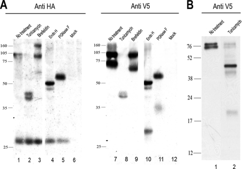
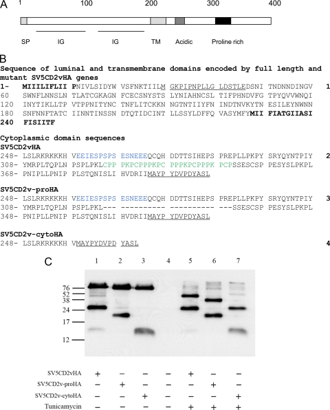
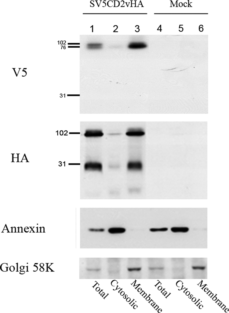
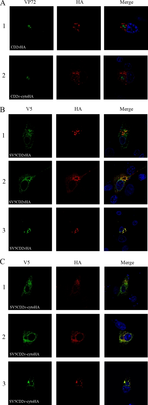
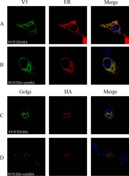
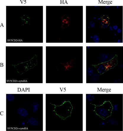
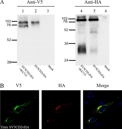
The African swine fever virus (ASFV)-encoded CD2v transmembrane protein is required for the hemadsorption of red blood cells around infected cells and is also required for the inhibition of bystander lymphocyte proliferation in response to mitogens. We studied the expression of CD2v by expressing the gene with a V5 tag downstream from the signal peptide near the N terminus and a hemagglutinin (HA) tag at the C terminus. In ASFV-infected cells, a full-length glycosylated form of the CD2v protein, which migrated mainly as a 89-kDa product, was detected, as well as an N-terminal glycosylated fragment of 63 kDa and a C-terminal nonglycosylated fragment of 26 kDa. All of these forms of the protein were localized in the membrane fraction of cells. The 26-kDa C-terminal fragment was also produced in infected cells treated with brefeldin A. These data indicate that the CD2v protein is cleaved within the luminal domain and that this occurs in the endoplasmic reticulum or Golgi compartments. Confocal microscopy showed that most of the expressed CD2v protein was localized within cells rather than at the cell surface. Comparison of the localization of full-length CD2v with that of a deletion mutant lacking all of the cytoplasmic tail apart from the 12 membrane-proximal amino acids indicated that signals within the cytoplasmic tail are responsible for the predominant localization of the full-length and C-terminal 26-kDa fragment within membranes around the virus factories, which contain markers for the Golgi compartment. Processing of the CD2v protein was not observed in uninfected cells, indicating that it is induced by ASFV infection.


Figures

FIG. 1.
FIG. 1.
Western blot showing CD2v protein expression in ASFV-infected Vero cells. Vero cells were transfected with plasmid SV5CD2vHA expressing the full-length EP402R (CD2v) protein tagged at the C terminus with the HA epitope tag and near the N terminus downstream from the signal peptide with the SV5 tag. The cells were then infected with the ASFV BA71V isolate and lysed at 18 h postinfection. (A) Lanes 1 to 6, proteins precipitated from lysates with anti-HA matrix, separated by SDS-PAGE, and blotted onto membranes. Immunoprecipitated proteins were detected by probing with anti-HA coupled to HRP, followed by enhanced chemiluminescence (ECL). In lanes 7 to 12, the lysates were immunoprecipitated using anti-V5 agarose, and the V5-tagged proteins were detected by probing Western blots of the immunoprecipitated proteins using anti-V5 coupled to HRP. Lanes 1 and 7, untreated lysates; lanes 2 and 8, lysates from cells treated with tunicamycin overnight at 1 μg ml−1 before being harvested; lanes 3 and 9, lysates from cells treated with brefeldin overnight at 15 μg ml−1 before being harvested. In lanes 4 and 10, cell lysates were digested with endo H prior to immunoprecipitation, and in lanes 5 and 11, the lysates were treated with PNGase F prior to immunoprecipitation. Lanes 6 and 12 show lysates from mock-transfected cells. (B) Cells transfected with a plasmid expressing SV5CD2vHA were infected with the ASFV BA71V isolate, and SV5-tagged proteins were detected as described above. Lane 1 shows untreated lysate, and lane 2 shows lysates from cells treated overnight with tunicamycin at 1 μg ml−1. The positions of molecular weight markers run in parallel are shown. The lanes shown in panel A were run on the same gel, and those in panel B were run on a different gel.
FIG. 2.
FIG. 2.
Western blot showing expression of mutant forms of CD2v. (A) Diagram indicating the locations of domains in the CD2v protein. The scale in amino acids is shown at the top. Domains indicated by boxes or underlined include the signal peptide (SP), immunoglobulin superfamily-like domains (IG), transmembrane domain (TM), acidic domain, and proline-rich domain. (B) Sequences (numbered on the right) of mutant forms of the CD2v protein. Sequence 1 is the sequence of the N-terminal 245 residues encoding the extracellular/luminal and transmembrane domains of the CD2v protein of the Malawi LIL 20/1 isolate. The sequence of the inserted SV5 tag is underlined. Sequence 1 is included in all of the mutant proteins. The signal peptide and transmembrane domains are in boldface. Sequences 2 to 4 are the sequences of the cytoplasmic domains of the full-length CD2v cytoplasmic tail, SV5CD2vHA (sequence 2); the deletion mutant lacking proline-rich repeats, SV5CD2v-proHA (sequence 3); and the deletion mutant lacking most of the cytoplasmic tail, SV5CD2v-cytoHA (sequence 4). The sequence of the C-terminal HA tag is underlined. Proline-rich-repeat sequences are in green, and acid-rich sequences are in blue. (C) Vero cells were transfected with plasmids expressing the full-length or deleted forms of the CD2v protein and infected with the ASFV BA71V isolate for 18 h. Cell lysates were immunoprecipitated using anti-HA matrix, and the immunoprecipitated proteins were separated by SDS-PAGE and blotted onto membranes. The blots were probed with anti-HA-HRP, and bound antibodies were detected by ECL. Lanes 1 and 5, extracts from cells expressing CD2vHA; lanes 2 and 6, extracts from cells expressing CD2v-proHA; lanes 3 and 7, extracts from cells expressing CD2v-cytoHA. Lanes 5, 6, and 7 show extracts from cells treated with tunicamycin (1 μg ml−1) overnight. Lane 4 shows extracts from mock-transfected Vero cells. The positions of molecular weight markers run in parallel are shown.
FIG. 3.
FIG. 3.
Western blot showing the localization of CD2v protein fragments in membrane or cytosolic cell fractions in ASFV-infected cells. Vero cells were transfected with plasmids expressing SV5CD2vHA and infected with the ASFV BA71V isolate. At 18 h postinfection, the cells were lysed and separated into cytosolic or membrane fractions. Lanes 1, 2, and 3, extracts from cells expressing the SV5CD2vHA protein; lanes 4, 5, and 6, extracts from mock-transfected cells. Lanes 1 and 4 show total cell extracts prepared by lysis in RIPA buffer, and lanes 2 to 6 show extracts fractionated into the cytosolic (lanes 2 and 5) and membrane (lanes 3 and 6) fractions. Extracts were separated by SDS-PAGE and blotted onto membranes. The blots were probed as indicated on the left with anti-V5-HRP, anti-HA-HRP, anti-annexin followed by anti-mouse-HRP, and anti-Golgi 58K protein followed by anti-mouse-HRP. Bound antibodies were detected by ECL.
FIG. 4.
FIG. 4.
Localization of CD2V in ASFV-infected cells. Vero cells were grown on coverslips and transfected with plasmids expressing full-length CD2v or deletion mutants and then infected with the ASFV BA71V isolate. The cells were fixed with paraformaldehyde at 18 h postinfection, permeabilized, and then stained with primary and secondary antibodies as indicated at the bottom and visualized by confocal microscopy. (A) Images of cells probed with anti-VP72 (green) and anti-HA (red), followed by appropriate Alexa Fluor-stained secondary antibodies. Row 1 shows cells expressing CD2vHA, and row 2 shows cells expressing CD2v-cytoHA. (B) Rows 1, 2, and 3 show different cells expressing SV5CD2vHA. (C) Rows 1, 2, and 3 show different cells expressing SV5CD2v-cytoHA. The SV5CD2vHA and SV5CD2v-cytoHA proteins were detected using anti-V5 tag (green) and anti-HA tag (red) antibodies, followed by the appropriate Alexa Fluor-conjugated secondary antibodies, and visualized by confocal microscopy. Merged images with DNA stained with DAPI are on the right.
FIG. 5.
FIG. 5.
Colocalization of SV5CD2vHA and SV5CD2v-cytoHA proteins within cellular compartments. Vero cells were grown on coverslips, transfected with plasmids expressing SV5CD2vHA or SV5CD2v-cytoHA, and infected with the BA71V ASFV isolate. The cells were fixed with paraformaldehyde at 18 h postinfection, permeabilized, stained with primary and secondary antibodies as indicated, and visualized by confocal microscopy. (A and C) Cells expressing SV5CD2vHA. (B and D) Cells expressing SV5CD2v-cytoHA. Panels A and B show staining with anti-V5 (green) and an anti-ERp60 marker (red). Panels C and D show staining with the anti-Golgi marker GM130 (green) and anti-HA (red), followed by appropriate Alexa Fluor-stained secondary antibodies. Merged images with DNA stained with DAPI are on the right.
FIG. 6.
FIG. 6.
Localization of cell surface and internalized SV5CD2vHA and SV5CD2v-cytoHA in ASFV-infected cells. Vero cells were grown on coverslips, transfected with plasmids expressing SV5CD2vHA or SV5CD2v-cytoHA, and then infected with the ASFV isolate BA71V. At 17 h postinfection, anti-V5 antibody was added to the medium (10-μg ml−1 final concentration), and incubation was continued for 1 h at 37°C. The cells were washed, fresh medium was added, and incubation continued at 37°C for 10 min before the cells were fixed with paraformaldehyde. (A and B) Cells transfected with SV5CD2vHA and SV5CD2v-cytoHA, respectively, that were permeabilized before being stained with Alexa Fluor-conjugated secondary antibody. These cells were also stained with anti-HA (red). (C) Cells transfected with SV5CD2v-cytoHA that were not permeabilized before being stained to detect the anti-V5 antibody. Merged images with DNA stained with DAPI are on the right.
FIG. 7.
FIG. 7.
Expression and localization of full-length CD2v in uninfected cells. (A) Vero cells were transfected with plasmid TriexSV5CD2vHA, and at 24 h posttransfection, lysates were prepared, or cells were transfected with plasmid SV5CD2vHA and infected with the ASFV BA71V isolate, and then lysates were prepared at 18 h postinfection. The lysates were immunoprecipitated using anti-HA matrix or anti-V5 agarose. The immunoprecipitates were separated by SDS-PAGE, transferred to membranes, and blotted using anti-HA antibody conjugated to HRP (lanes 1, 2, and 3) or anti-V5 antibody conjugated to HRP (lanes 4, 5, and 6). Lanes 1 and 4, lysates from infected cells expressing SV5CD2vHA; lanes 2 and 5, lysates from uninfected cells transfected with plasmid TriexSV5CD2vHA; lanes 3 and 6, lysates from mock-transfected cells. The positions of molecular weight markers run in parallel are indicated. (B) Cells were grown on coverslips, transfected with plasmid TriexSV5CD2vHA, and then fixed with paraformaldehyde at 24 h posttransfection. The cells were permeabilized and stained with anti-V5 antibody (green) and anti-HA antibody (red), followed by appropriate Alexa Fluor-conjugated secondary antibodies, and then visualized by confocal microscopy. A merged image with DNA stained with DAPI is on the right.

Similar articles

Cited by

References

    1. Arias, M., and J. M. Sanchez-Vizcaino. 2002. African swine fever, p. 119-124. In A. Morilla Gonzaìlez, K.-J. Yoon, and J. J. Zimmerman (ed.), Trends in emerging viral infections of swine. Iowa State Press, Ames, IA.
    1. Barclay, A. N. 2001. Biochemical analysis of the lymphocyte cell surface—from alloantisera to the role of membrane proteins. Immunol. Rev. 184:69-81. - PubMed
    1. Bodian, D. L., E. Y. Jones, K. Harlos, D. I. Stuart, and S. J. Davis. 1994. Crystal structure of the extracellular region of the human cell-adhesion molecule cd2 at 2.5-Angstrom resolution. Structure 2:755-766. - PubMed
    1. Boinas, F. S., G. H. Hutchings, L. K. Dixon, and P. J. Wilkinson. 2004. Characterization of pathogenic and non-pathogenic African swine fever virus isolates from Ornithodoros erraticus inhabiting pig premises in Portugal. J. Gen. Virol. 85:2177-2187. - PubMed
    1. Borca, M. V., et al. 1998. Deletion of a CD2-like gene, 8-DR, from African swine fever virus affects viral infection in domestic swine. J. Virol. 72:2881-2889. - PMC - PubMed

Publication types

MeSH terms

LinkOut - more resources