. 2011 Apr;85(7):3415-23.
doi: 10.1128/JVI.01768-10. Epub 2011 Jan 19.

Vital role for CD8+ cells in controlling retroviral infections

Affiliations

Vital role for CD8+ cells in controlling retroviral infections

Melissa Kane et al. J Virol. 2011 Apr.

Abstract

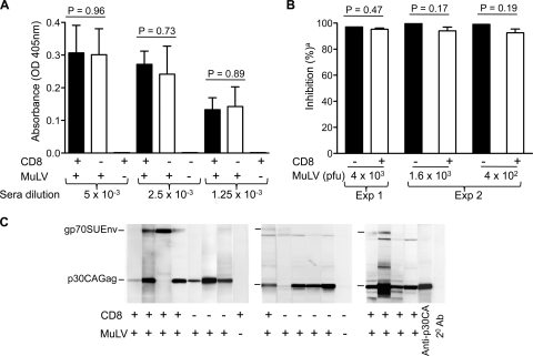
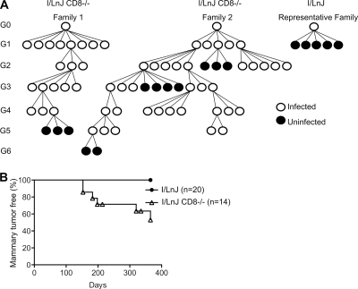
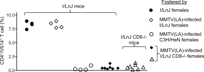
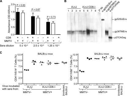
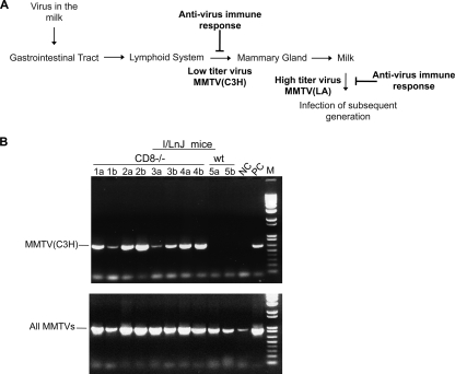
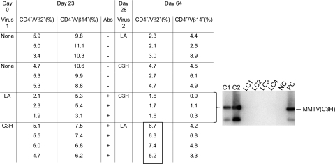
Antiviral adaptive immune defenses consist of humoral and cell-mediated responses, which together eliminate extracellular and intracellular virus. As most retrovirus-infected individuals do not raise efficient protective antivirus immune responses, the relative importance of humoral and cell-mediated responses in restraining retroviral infection is not well understood. We utilized retrovirus-resistant I/LnJ mice, which control infection with mouse mammary tumor virus (MMTV) and murine leukemia virus (MuLV) via an adaptive immune mechanism, to assess the contribution of cellular responses and virus-neutralizing antibodies (Abs) to the control of retroviral infection. We found that in retrovirus-infected CD8-deficient I/LnJ mice, viral titers exceed the neutralizing capability of antiviral Abs, resulting in augmented virus spread and disease induction. Thus, even in the presence of robust neutralizing Ab responses, CD8-mediated responses are essential for full protection against retroviral infection.


Figures

FIG. 1.
FIG. 1.
MuLV-infected CD8-deficient I/LnJ mice produce antivirus neutralizing Abs. (A) MuLV-infected CD8−/− and CD8+/+ I/LnJ mice produce antivirus Abs of similar titers. CD8−/− (n = 4) and CD8+/+ (n = 6) I/LnJ mice were infected with MuLV, and their sera were tested for reactivity against MuLV virion proteins in an ELISA at 8 weeks postinfection. Sera from uninfected CD8−/− and CD8+/+ I/LnJ mice were used as controls. The results for three different dilutions of sera are shown. The antiretroviral Ab response in I/LnJ mice is IFN-γ mediated and is predominantly of the IgG2a isotype (47). Therefore, anti-mouse IgG2a-specific Abs coupled to AP were used at the second step. OD 405, optical density at 405 nm. An unpaired t test was applied to determine the P value. (B) Abs produced by MuLV-infected CD8−/− I/LnJ mice are virus neutralizing. MuLV virions were incubated with sera of uninfected I/LnJ mice (pool of three serum specimens) and MuLV-infected I/LnJ (pool of three serum specimens) or I/LnJ CD8−/− (n = 7) mice and then subjected to an XC plaque assay. a, percent inhibition relative to that by uninfected sera (no inhibition). Exp 1 and 2, two different experiments performed with distinct serum specimens. Sera from four and three MuLV-infected CD8−/− I/LnJ mice were used in experiment 1 and experiment 2, respectively. Reactivity of the mouse sera with viral proteins is shown in panel C. An unpaired t test was applied to determine the P value. (C) Antivirus Abs produced by MuLV-infected CD8−/− I/LnJ mice recognize virion proteins. PVDF strips with virion proteins separated on an SDS-polyacrylamide gel were incubated with the indicated mouse sera (which were also used for neutralization studies). anti-p30CA, rat monoclonal Ab against p30CAGag. Anti-mouse IgG2a-specific and anti-rat Ig-specific secondary (2° Ab) Abs coupled to HRP were used at the second step. gp70SUEnv, surface glycoprotein, product of the env gene; p30CAGag, capsid protein, product of the gag gene.
FIG. 2.
FIG. 2.
CD8+ cells are required for efficient elimination of MMTV in vivo and to resist virally induced mammary tumors. (A) Scheme showing virus fate in MMTV(LA)-infected CD8−/− and control CD8+/+ I/LnJ mouse pedigrees. G0 mice were fostered on MMTV(LA)-infected C3H/HeN females. Animals were monitored for infection by analyzing the percentages of SAg-cognate T cells in the periphery. Results for 1 of over 30 representative families of control I/LnJ mice are depicted. Circles represent individual mice (male or female). Open circles, virus-infected mice; filled circles, virus-free mice. (B) Mammary gland tumor development in MMTV(LA)-infected CD8−/− and CD8+/+ I/LnJ mice. MMTV(LA)-infected CD8−/− (only females that showed deletion of SAg-cognate T cells from G0 to G3 were used for the studies) and CD8+/+ I/LnJ (G0) females were bred and monitored for mammary tumors for 365 days.
FIG. 3.
FIG. 3.
Transmission of infectious MMTV by CD8-deficient I/LnJ mice. I/LnJ mice were fostered by uninfected I/LnJ, MMTV(LA)-infected I/LnJ, MMTV(LA)-infected C3H/HeN, or MMTV(LA)-infected CD8−/− I/LnJ females. Results for CD8−/− I/LnJ mice fostered by MMTV(LA)-infected CD8−/− I/LnJ mice are shown as a control. Frequencies of peripheral Vβ14+ cells among CD4+ T cells in infected mice were analyzed at 8 to 10 weeks of age. Each symbol represents an individual mouse.
FIG. 4.
FIG. 4.
MMTV-infected CD8-deficient I/LnJ mice produce virus-neutralizing Abs. (A) MMTV(LA)-infected CD8−/− and CD8+/+ I/LnJ mice produce similar titers of antivirus Abs. MMTV(LA)-infected (via fostering) CD8−/− (n = 8), CD8+/+ I/LnJ (n = 5), and uninfected I/LnJ (n = 5) mice were tested for production of Abs reactive against MMTV virion proteins by ELISA at 12 weeks of age. Anti-mouse IgG2a-specific Abs coupled to AP were used at the second step. Three different dilutions of sera are shown. OD 405, optical density at 405 nm. Unpaired t test was applied to determine the P value. (B) MMTV-infected CD8−/− I/LnJ mice produce Abs that recognize virion proteins. PVDF strips with virion proteins separated on an SDS-gel were incubated with mouse MAbs against capsid Gag (αp27CAGag), transmembrane Env (αgp36TMEnv), surface Env (αgp52SUEnv), or the indicated mouse sera. Anti-mouse polyisotypic (for MAbs) or IgG2a-specific (for mouse sera) Abs coupled to HRP were used at the second step. Numbers represent individual mice. The vast majority of Abs observed by Western blotting are directed against gp36TMEnv (; this study). (C) Abs produced by MMTV(LA)-infected CD8-deficient I/LnJ mice are capable of neutralizing the virus in vitro. MMTV(LA) virions were incubated with sera from MMTV(LA)-infected CD8−/− I/LnJ, control I/LnJ mice, or uninfected I/LnJ mice and injected i.p. into virus-susceptible BALB/cJ mice. At 4 weeks postinjection, frequencies of peripheral Vβ6+, Vβ14+, and Vβ2+ cells (data not shown) among CD4+ T cells in the periphery were analyzed by FACS analysis. Each symbol represents an individual mouse. Uninfected BALB/cJ, percentages of CD4+/Vβ14+ and CD4+/Vβ6+ among CD4+ T cells in the periphery of uninfected BALB/cJ mice.
FIG. 5.
FIG. 5.
Viral infection with a low-titer virus progresses to the mammary glands in the absence of CD8+ cells. (A) Scheme representing the MMTV infectious cycle and phenotypes of I/LnJ mice infected with different virus variants. Antiviral immune responses in I/LnJ mice infected with the low-titer variant MMTV(C3H) prevent viral progression from the lymphoid system to the mammary glands, while in I/LnJ mice infected with the high-titer MMTV(LA), virus progresses to the mammary gland and is secreted into the milk (10, 18, 47). Secreted virus, however, is coated with Abs and is thus unable to establish infection in the offspring. (B) MMTV(C3H) progresses to the mammary gland of CD8-deficient I/LnJ mice. RNA isolated from the lactating mammary gland and the milk of CD8−/− or CD8+/+ I/LnJ mice fostered by MMTV(C3H)-infected mothers was subjected to RT-PCR analysis specific for MMTV(C3H) (top) or for all MMTVs (bottom) (milk contains cells shed by the mammary glands, which express endogenous proviruses). Lanes: numbers, individual mice; a, RNA isolated from lactating mammary glands; b, RNA isolated from milk; NC, negative control (RNA isolated from the milk of an uninfected I/LnJ mouse); PC, positive control (DNA isolated from mammary gland tumors of C3H/HeN mice infected with MMTV(C3H); M, molecular size marker, 1-kb ladder (Invitrogen, Carlsbad, CA).
FIG. 6.
FIG. 6.
Antiviral immune responses in MMTV-infected I/LnJ mice are sufficient to prevent superinfection with distinct viruses. I/LnJ mice were infected by i.p. injection with MMTV(C3H) or MMTV(LA). Twenty-three days later, frequencies of peripheral Vβ2+ and V14+ cells among CD4+ T cells and antiviral Ab production were examined by FACS analysis and ELISA, respectively. Mice were i.p. injected with the opposite viral variant [control mice were injected with either MMTV(C3H) or MMTV(LA)] on day 28. Thirty-six days following the final injection, mice initially infected with MMTV(C3H) were screened for evidence of the second infection with MMTV(LA) by FACS analysis. The boxed percentages of cells are indistinguishable from the percentages of the same cells in uninfected mice (P = 0.08), indicating that MMTV(LA)-superinfected mice were not infected with the virus. As both MMTV(C3H) and MMTV(LA) variants delete Vβ14+-cognate T cells, superinfection of MMTV(LA)-infected mice with MMTV(C3H) was monitored by examining the spleens for the presence of integrated MMTV(C3H) provirus. While mice singly infected with MMTV(C3H) contain newly integrated provirus in their spleens, MMTV(C3H) provirus was absent in MMTV(C3H)-superinfected mice. CD4+/Vβ2(14)+ (%), percentage of Vβ2+ or Vβ14+ cells among CD4+ T cells. Lanes: C1 and C2, DNA isolated from spleens of I/LnJ mice injected with MMTV(C3H) on day 28; LC1, LC2, LC3, and LC4, DNA isolated from spleens of I/LnJ mice injected with MMTV(LA) on day 0 and MMTV(C3H) on day 28; NC, negative control (splenic DNA isolated from an uninfected I/LnJ mouse); PC, positive control [DNA isolated from mammary gland tumor of a MMTV(C3H)-infected C3H/HeN mouse].

Similar articles

Cited by

References

    1. Arvin, A. 1998. Varicella-zoster virus: virologic and immunologic aspects of persistent infection, p. 183-208. In R. Ahmed and I. Chen (ed.), Persistent viral infections. John Wiley & Sons, Inc., New York, NY.
    1. Bailey, J. R., et al. 2006. Neutralizing antibodies do not mediate suppression of human immunodeficiency virus type 1 in elite suppressors or selection of plasma virus variants in patients on highly active antiretroviral therapy. J. Virol. 80:4758-4770. - PMC - PubMed
    1. Betts, M. R., et al. 2006. HIV nonprogressors preferentially maintain highly functional HIV-specific CD8+ T cells. Blood 107:4781-4789. - PMC - PubMed
    1. Binder, G. K., and D. E. Griffin. 2003. Immune-mediated clearance of virus from the central nervous system. Microbes Infect. 5:439-448. - PubMed
    1. Boeke, J. D., and J. P. Stoye. 1997. Retrotransposons, endogenous retroviruses, and the evolution, p. 343-436. In J. M. Coffin, S. H. Hughes, and H. E. Varmus (ed.), Retroviruses. Cold Spring Harbor Laboratory Press, Cold Spring Harbor, NY. - PubMed

Publication types

MeSH terms