. 2011 Apr;85(7):3095-105.
doi: 10.1128/JVI.02360-10. Epub 2011 Jan 19.

Rotavirus VP2 core shell regions critical for viral polymerase activation

Affiliations

Rotavirus VP2 core shell regions critical for viral polymerase activation

Sarah M McDonald et al. J Virol. 2011 Apr.

Abstract

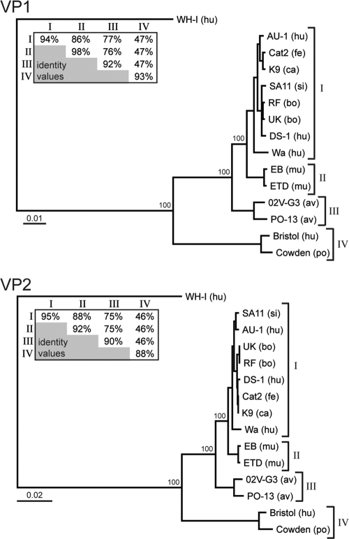
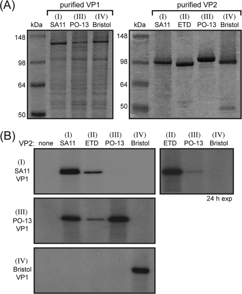
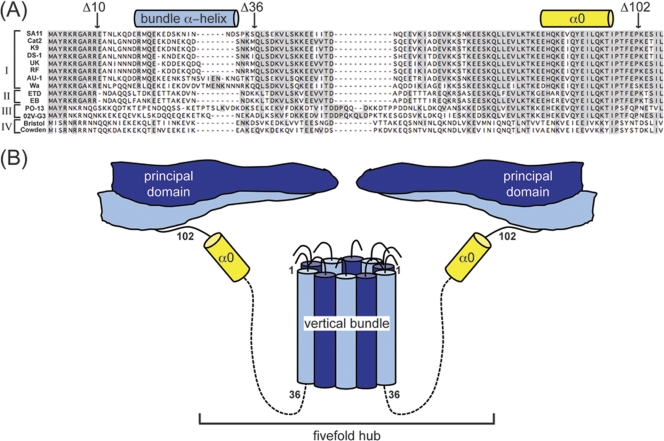
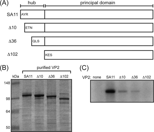
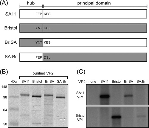
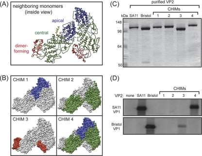
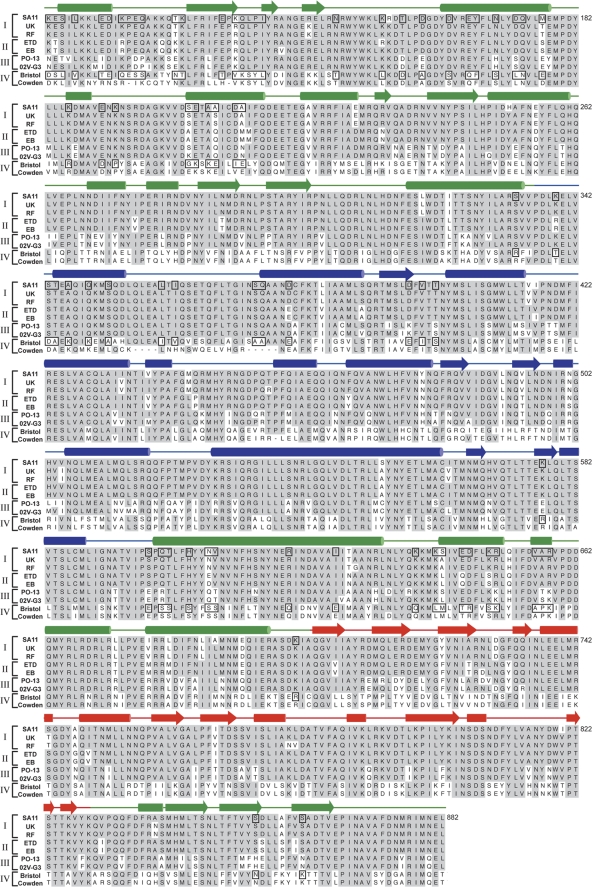
The innermost VP2 core shell of the triple-layered, icosahedral rotavirus particle surrounds the viral genome and RNA processing enzymes, including the RNA-dependent RNA polymerase (VP1). In addition to anchoring VP1 within the core, VP2 is also an essential cofactor that triggers the polymerase to initiate double-stranded RNA (dsRNA) synthesis using packaged plus-strand RNA templates. The VP2 requirement effectively couples packaging with genome replication and ensures that VP1 makes dsRNA only within an assembling previrion particle. However, the mechanism by which the rotavirus core shell protein activates the viral polymerase remains very poorly understood. In the current study, we sought to elucidate VP2 regions critical for VP1-mediated in vitro dsRNA synthesis. By comparing the functions of proteins from several different rotaviruses, we found that polymerase activation by the core shell protein is specific. Through truncation and chimera mutagenesis, we demonstrate that the VP2 amino terminus, which forms a decameric, internal hub underneath each 5-fold axis, plays an important but nonspecific role in VP1 activation. Our results indicate that the VP2 residues correlating with polymerase activation specificity are located on the inner face of the core shell, distinct from the amino terminus. Based on these findings, we predict that several regions of VP2 engage the polymerase during the concerted processes of rotavirus core assembly and genome replication.


Figures

FIG. 1.
FIG. 1.
Structure of the rotavirus VP2 core shell. (Left) Structure of the T=1 icosahedral core shell of bovine rotavirus (PDB accession number 3KZ4), with each of the 120 VP2 monomers depicted in a surface representation. Five type A and five type B VP2 monomers of a central decamer are in dark blue and light blue, respectively. (Right) Inside view of two neighboring VP2 A-B dimers. Type A and B monomers are in dark blue and light blue, respectively. One of the dimers (comprised of monomers A1 and B1) is depicted in a ribbon representation, while the other (comprised of monomers A2 and B2) is depicted in a surface representation. The resolved portion of the VP2 amino terminus in type B monomers (residues 81 to 100) is yellow and is shown in a ribbon representation.
FIG. 2.
FIG. 2.
Genetic divergence of VP1 and VP2 from different rotavirus strains. Phylogenetic dendrograms of VP1 (top) or VP2 (bottom) were constructed by using the amino acid sequences and the neighbor-joining method (1,000 bootstrap repetitions). The virus strain is listed with the species of isolation in parentheses: human (hu), simian (si), bovine (bo), porcine (po), feline (fe), canine (ca), murine (mu), and avian (av). The proteins of a group B rotavirus (strain WH-1) were used to root the trees. Bootstrap values are shown as percentages for key nodes. Brackets indicate the designated category (category I, II, III, or IV) of the VP1 and VP2 proteins. The average intra- and intercategory percent amino acid identities (identity values) are shown in the inset tables. The scale bar represents the number of substitutions per amino acid position.
FIG. 3.
FIG. 3.
Functional compatibility of VP1 and VP2 proteins. (A) Purified VP1 and VP2 proteins. Recombinant proteins were expressed in insect cells using baculovirus vectors and purified as described in Materials and Methods. The designated category (category I, II, III, or IV) of each protein is shown in parentheses above the strain. Samples containing approximately 2 pmol of VP1 or 20 pmol of VP2 were electrophoresed in a 10% SDS-polyacrylamide gel and visualized by PageBlue staining. Molecular mass markers are shown (in kilodaltons). (B) In vitro dsRNA synthesis by SA11 (top), PO-13 (middle), or Bristol (bottom) VP1. The designated category (category I, III, or IV) of each polymerase is shown in parentheses above the strain. Reactions proceeded in the absence (none) or presence of the different core shell proteins listed above the gels. Radiolabeled dsRNA products were resolved with 10% SDS-polyacrylamide gels and detected by autoradiography. The images on the left represent 4-h exposures (exp) of the gels to film. To visualize SA11 VP1 activity in the presence of PO-13 VP2, the gel was exposed to film for 24 h.
FIG. 4.
FIG. 4.
Sequence and predicted architecture of the VP2 5-fold hub. (A) Alignment of the rotavirus VP2 amino termini. The VP2 amino acid sequences of several representative virus strains are shown. Dashes indicate gaps in the protein sequence, and shading reveals the conservation of amino acid identity. Brackets indicate the designated category (category I, II, III, or IV) of the VP2 proteins based on phylogenetics. The residues comprising the bundle α-helix (light blue cylinder) and the first visible helix in the crystal structure (α0) (yellow cylinder) are shown schematically, as are the sites of truncations for the experiments described in the legend of Fig. 5. (B) Cartoon rendering of the VP2 5-fold hub. Type A and B monomers are shown in dark blue and light blue, respectively. The VP2 amino termini (residues ∼1 to 100) of 10 abutting monomers in a decamer unit oligomerize underneath the 5-fold axis. Residues 18 to 31 form an α-helix that clusters with those of neighboring monomers to create a 10-helix vertical bundle. Only the principal domains of two dimers and the hub connections of two monomers are shown for simplicity. The cartoon is meant to roughly illustrate the approximate shape and location of the 5-fold hub, the vertical bundle, α0 (yellow), and the principle domain (comprised of residues ∼100 to 880) and is not drawn to scale.
FIG. 5.
FIG. 5.
Truncation mutagenesis of the SA11 VP2 5-fold hub. (A) Cartoon schematics of wild-type and mutant SA11 VP2 proteins. The residues contributing to the 5-fold hub are delineated from those of the principal domain by a black line. The three amino acids that follow the starting methionine are listed for the wild type (SA11) and each mutant protein (Δ10, Δ36, and Δ102). (B) Purified VP2 proteins. VP2 proteins were electrophoresed in a 10% SDS-polyacrylamide gel and visualized by PageBlue staining. Molecular size markers are shown (in kilodaltons). (C) In vitro dsRNA synthesis by SA11 VP1. Reactions proceeded in the absence (none) or the presence of the different core shell proteins listed above the gel. Radiolabeled dsRNA products were resolved with 10% SDS-polyacrylamide gels and detected by autoradiography.
FIG. 6.
FIG. 6.
Fivefold hub chimeric VP2 proteins. (A) Cartoon schematics of wild-type and chimeric VP2 proteins. Wild-type SA11 and Bristol VP2 proteins are shown in white and gray, respectively. The residues contributing to the 5-fold hub are delineated from those of the principal domain by a black line. Schematics of the 5-fold hub chimeras (Br:SA and SA:Br) are shown, with the parental origin of amino acids indicated by color (white for SA11 or gray for Bristol). The three amino acids before and after the fusion site are listed. (B) Purified VP2 proteins. VP2 proteins were electrophoresed in a 10% SDS-polyacrylamide gel and visualized by PageBlue staining. Molecular size markers are shown (kilodaltons). (C) In vitro dsRNA synthesis by SA11 or Bristol VP1. Reactions proceeded in the absence (none) or the presence of the different core shell proteins listed above the gels. Radiolabeled dsRNA products were resolved with 10% SDS-polyacrylamide gels and detected by autoradiography.
FIG. 7.
FIG. 7.
Subdomain chimeric VP2 proteins. (A) Ribbon representation of neighboring VP2 monomers in a dimeric unit (inside view). The apical, central, and dimer-forming subdomains are in blue, green, and red, respectively. The resolved portion of the VP2 amino terminus in the type B monomer (residues 81 to 100) is in yellow. (B) Surface representations of chimeric (CHIM) VP2 proteins. In all images, gray indicates Bristol VP2 residues, while color indicates SA11 VP2 residues. (C) Purified VP2 proteins. VP2 proteins were electrophoresed in a 10% SDS-polyacrylamide gel and visualized by PageBlue staining. Molecular size markers are shown (in kilodaltons). (D) In vitro dsRNA synthesis by SA11 or Bristol VP1. Reactions proceeded in the absence (none) or the presence of the different core shell proteins listed above the gel. Radiolabeled dsRNA products were resolved with 10% SDS-polyacrylamide gels and detected by autoradiography.
FIG. 8.
FIG. 8.
Alignment of the VP2 principal domain. The VP2 amino acid sequences of several representative virus strains are shown. Dashes indicate gaps in the protein alignment, and shading reveals the conservation of amino acid identity. Brackets indicate the designated category (category I, II, III, or IV) of the VP2 proteins based on the phylogenetic analyses described in the legend of Fig. 2. Subdomains are colored as described in the legend of Fig. 7, and secondary structural elements are shown above the corresponding residues (12). Amino acids outlined in black were targeted for multipoint (MP) mutagenesis in Fig. 9.
FIG. 9.
FIG. 9.
Multipoint mutagenesis of Bristol VP2. (A) Three-dimensional location of amino acids identified in Fig. 8 (outlined residues) on the inner surface of the core shell. Neighboring VP2 monomers in a dimeric unit (inside view) are seen in a surface representation. Point mutations of residues of the apical (api) and central (cent) subdomains are shown in blue and green, respectively. Each Bristol VP2 residue at the colored sites was changed to match the corresponding residue of SA11 VP2. (B) Purified VP2 proteins. VP2 proteins were electrophoresed in a 10% SDS-polyacrylamide gel and visualized by PageBlue staining. Molecular size markers are shown (in kilodaltons). (C) In vitro dsRNA synthesis. Reactions proceeded in the absence (none) or the presence of the different core shell proteins listed above the gel. Radiolabeled dsRNA products were resolved with 10% SDS-polyacrylamide gels and detected by autoradiography.
FIG. 10.
FIG. 10.
Cartoon model of the VP1-VP2 interaction(s) that leads to polymerase activation. The images show cartoon renderings of a VP2 decamer interacting with a VP1 monomer. Type A and B monomers are in dark blue and light blue, respectively. Residues 1 to 81 of type B monomers are illustrated as yellow rectangles. VP1 (gray) is shown in relative scale binding to the VP2 core shell principal domain and juxtaposed to the 5-fold hub. (A) Inside view. The approximate scaled diameter of the hub is indicated as a black dotted line. (B) Side view as described in the legend of Fig. 4.

Similar articles

Cited by

References

    1. Arnoldi, F., M. Campagna, C. Eichwald, U. Desselberger, and O. R. Burrone. 2007. Interaction of rotavirus polymerase VP1 with nonstructural protein NSP5 is stronger that that with NSP2. J. Virol. 81:2128-2137. - PMC - PubMed
    1. Berois, M., C. Sapin, I. Erk, D. Poncet, and J. Cohen. 2003. Rotavirus nonstructural protein NSP5 interacts with major core protein VP2. J. Virol. 77:1757-1763. - PMC - PubMed
    1. Estes, M. K., and A. Z. Kapikian. 2007. Rotaviruses and their replication, p. 1917-1974. In D. M. Knipe et al. (ed.), Fields virology, 5th ed. Lippincott Williams & Wilkins, Philadelphia, PA.
    1. Gallegos, C. O., and J. T. Patton. 1989. Characterization of rotavirus replication intermediates: a model for the assembly of single-shelled particles. Virology 172:616-627. - PubMed
    1. Guglielmi, K. M., S. M. McDonald, and J. T. Patton. 2010. Mechanism of intra-particle synthesis of the rotavirus double-stranded RNA genome. J. Biol. Chem. 285:18123-18128. - PMC - PubMed

Publication types

MeSH terms

Associated data

LinkOut - more resources