. 2011 May;300(5):C1023-33.
doi: 10.1152/ajpcell.00288.2010. Epub 2011 Jan 19.

Small GTPase Rab11b regulates degradation of surface membrane L-type Cav1.2 channels

Affiliations

Small GTPase Rab11b regulates degradation of surface membrane L-type Cav1.2 channels

Jabe M Best et al. Am J Physiol Cell Physiol. 2011 May.

Abstract

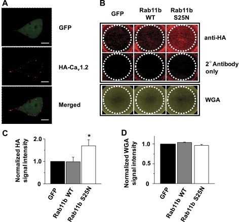
L-type Ca(2+) channels (LTCCs) play a critical role in Ca(2+)-dependent signaling processes in a variety of cell types. The number of functional LTCCs at the plasma membrane strongly influences the strength and duration of Ca(2+) signals. Recent studies demonstrated that endosomal trafficking provides a mechanism for dynamic changes in LTCC surface membrane density. The purpose of the current study was to determine whether the small GTPase Rab11b, a known regulator of endosomal recycling, impacts plasmalemmal expression of Ca(v)1.2 LTCCs. Disruption of endogenous Rab11b function with a dominant negative Rab11b S25N mutant led to a significant 64% increase in peak L-type Ba(2+) current (I(Ba,L)) in human embryonic kidney (HEK)293 cells. Short-hairpin RNA (shRNA)-mediated knockdown of Rab11b also significantly increased peak I(Ba,L) by 66% compared when with cells transfected with control shRNA, whereas knockdown of Rab11a did not impact I(Ba,L). Rab11b S25N led to a 1.7-fold increase in plasma membrane density of hemagglutinin epitope-tagged Ca(v)1.2 expressed in HEK293 cells. Cell surface biotinylation experiments demonstrated that Rab11b S25N does not significantly impact anterograde trafficking of LTCCs to the surface membrane but rather slows degradation of plasmalemmal Ca(v)1.2 channels. We further demonstrated Rab11b expression in ventricular myocardium and showed that Rab11b S25N significantly increases peak I(Ba,L) by 98% in neonatal mouse cardiac myocytes. These findings reveal a novel role for Rab11b in limiting, rather than promoting, the plasma membrane expression of Ca(v)1.2 LTCCs in contrast to its effects on other ion channels including human ether-a-go-go-related gene (hERG) K(+) channels and cystic fibrosis transmembrane conductance regulator. This suggests Rab11b differentially regulates the trafficking of distinct cargo and extends our understanding of how endosomal transport impacts the functional expression of LTCCs.


Figures

Fig. 1.
Fig. 1.
Disruption of endogenous Rab11b function with dominant negative Rab11b S25N mutant increases L-type Ba2+ current (IBa,L). A: representative IBa,L traces recorded from human embryonic kidney (HEK)293 cells transiently expressing wild-type (WT) Cav1.2, β2cN4, and either green fluorescent protein (GFP), Rab11b WT, or Rab11b S25N using whole cell rupture patch-clamp technique. B: mean current-voltage (I-V) relations constructed from GFP (n = 14), Rab11b WT (n = 4), and Rab11b S25N (n = 12) expressing cells. C: representative IBa,L traces from HEK293 cells transfected with WT Cav1.2, β2cN4, and either GFP, Rab11b WT, GTPase deficient Rab11b Q70L, or Rab11b S25N. D: mean I-V relations from GFP (n = 6), Rab11b WT (n = 5), Rab11b Q70L (n = 8), and Rab11b S25N (n = 8) expressing cells. *P < 0.05.
Fig. 2.
Fig. 2.
Short-hair pin RNA (shRNA)-mediated depletion of Rab11b increases IBa,L. A: Western blot analysis of HEK293 cells transfected with control shRNA or shRNA specific for Rab11b or Rab11a. To assess the relative knockdown of Rab11 isoforms, densitometric analysis was performed. Rab11b (B, n = 3) or Rab11a (C, n = 4) levels were normalized to GAPDH and compared with control shRNA. D: IBa,L recordings from HEK293 cells transfected with Cav1.2, β2cN4, and either control shRNA, Rab11b shRNA, or Rab11a shRNA. E: mean I-V relations constructed from control shRNA (n = 12), Rab11b shRNA (n = 13), and Rab11a shRNA (n = 11) expressing cells. *P < 0.05.
Fig. 3.
Fig. 3.
Rab11b S25N increases plasmalemmal expression of Cav1.2. A: confocal image of a nonpermeabilized HEK293 cell transiently transfected with HA-Cav1.2 (containing extracellular HA epitope), β2cN4, and GFP and immunolabeled with anti-hemagglutinin (HA) to visualize surface membrane Cav1.2 channels. White scale bar represents 5 μm. B: representative image of the infrared emission signal obtained from cells grown in a 12-well tissue culture plate. Top row: cells that were immunolabeled with anti-HA; middle row: primary antibody was omitted to depict background infrared signal. Bottom row: cells that were labeled with wheat germ agglutinin (WGA) Alexa Fluor 680 to control for cell density. White dotted circles represent regions of interest used for quantitative analysis. C: mean HA infrared signal intensity (n = 4) in which data were normalized to the GFP control group; *P < 0.05. D: mean WGA infrared signal intensity (n = 4) after normalizing to GFP control group.
Fig. 4.
Fig. 4.
Rab11b S25N increases plasmalemmal expression of Cav1.2 at late, but not early, time points after transient transfection. A: Western blot analysis of cell surface biotinylation studies performed 12, 24, or 36 h posttransfection. Samples were immunoprecipitated with anti-HA antibody and detected by immunoblotting with anti-HA (top) and anti-biotin (bottom). Average densitometric analysis (n = 4) of HA (B) and biotin (C) signal intensities over time for GFP (solid bars), Rab11b WT (shaded bars), and Rab11b S25N (open bars) expressing cells. HA and biotin signal intensities were normalized to their respective values from the GFP control group at 24 h posttransfection. *P < 0.05.
Fig. 5.
Fig. 5.
Slowed degradation of plasmalemmal Cav1.2 protein by Rab11b S25N. HEK293 cells were pulse-labeled with biotin and then incubated at 37°C + 5% CO2 for 0, 1, 3, or 6 h. Cell lysates were used for IP with anti-HA or incubated with NeutrAvidin and analyzed by SDS/PAGE and Western blotting. A: representative Western blot for total HA-Cav1.2 (top), biotin pulse-labeled HA-Cav1.2 (middle), and biotin-pulse labeled transferrin receptor (Tfn-R, bottom). Densitometric analysis (n = 3–6) was performed to quantitate total HA-Cav1.2 (B), biotin pulse-labeled HA-Cav1.2 (C), and biotin-pulse labeled Tfn-R (D) over time for GFP (■, solid line), Rab11b WT (●, dashed line), and Rab11b S25N (▲, dotted line) groups. Within each group, values were normalized to the signal intensity at t = 0. *P < 0.05.
Fig. 6.
Fig. 6.
Rab11b is expressed in ventricular myocyardium and regulates IBa,L in cardiomyocytes. A: RT-PCR analysis using RNA extracted from adult mouse brain tissue or freshly isolated left ventricular (LV) myocytes. B: Western blot analysis of adult mouse brain or LV homogenate using antibody specific for Rab11b. C: representative IBa,L recordings from neonatal mouse ventricular myocytes. D: mean I-V relations from myocytes transfected with GFP (n = 5), Rab11b WT (n = 6), and Rab11b S25N (n = 4). *P < 0.05.

References

    1. Altier C, Dubel SJ, Barrère C, Jarvis SE, Stotz SC, Spaetgens RL, Scott JD, Cornet V, De Waard M, Zamponi GW, Nargeot J, Bourinet E. Trafficking of L-type calcium channels mediated by the postsynaptic scaffolding protein AKAP79. J Biol Chem 277: 33598–33603, 2002 - PubMed
    1. Anderson CL, Delisle BP, Anson BD, Kilby JA, Will ML, Tester DJ, Gong Q, Zhou Z, Ackerman MJ, January CT. Most LQT2 mutations reduce Kv11.1 (hERG) current by a class 2 (trafficking-deficient) mechanism. Circulation 113: 365–373, 2006 - PubMed
    1. Antzelevitch C, Pollevick GD, Cordeiro JM, Casis O, Sanguinetti MC, Aizawa Y, Guerchicoff A, Pfeiffer R, Oliva A, Wollnik B, Gelber P, Bonaros EP, Jr, Burashnikov E, Wu Y, Sargent JD, Schickel S, Oberheiden R, Bhatia A, Hsu LF, Haïssaguerre M, Schimpf R, Borggrefe M, Wolpert C. Loss-of-function mutations in the cardiac calcium channel underlie a new clinical entity characterized by ST-segment elevation, short QT intervals, and sudden cardiac death. Circulation 115: 442–449, 2007 - PMC - PubMed
    1. Balijepalli RC, Foell JD, Hall DD, Hell JW, Kamp TJ. Localization of cardiac L-type Ca(2+) channels to a caveolar macromolecular signaling complex is required for beta(2)-adrenergic regulation. Proc Natl Acad Sci USA 103: 7500–7505, 2006 - PMC - PubMed
    1. Bers DM. Cardiac excitation-contraction coupling. Nature 415: 198–205, 2002 - PubMed

Publication types

MeSH terms

LinkOut - more resources