. 2011 Jan 19;31(3):809-18.
doi: 10.1523/JNEUROSCI.4330-10.2011.

MicroRNA miR-9 modifies motor neuron columns by a tuning regulation of FoxP1 levels in developing spinal cords

Affiliations

MicroRNA miR-9 modifies motor neuron columns by a tuning regulation of FoxP1 levels in developing spinal cords

Gaizka Otaegi et al. J Neurosci. .

Abstract

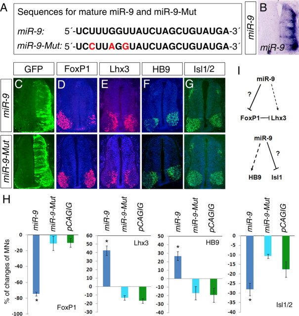
The precise organization of motor neuron subtypes in a columnar pattern in developing spinal cords is controlled by cross-interactions of multiple transcription factors and segmental expressions of Hox genes and their accessory proteins. Accurate expression levels and domains of these regulators are essential for organizing spinal motor neuron columns and axonal projections to target muscles. Here, we show that microRNA miR-9 is transiently expressed in a motor neuron subtype and displays overlapping expression with its target gene FoxP1. Overexpression or knockdown of miR-9 alters motor neuron subtypes, switches columnar identities, and changes axonal innervations in developing chick spinal cords. miR-9 modifies spinal columnar organization by specifically regulating FoxP1 protein levels, which in turn determine distinct motor neuron subtypes. Our findings demonstrate that miR-9 is an essential regulator of motor neuron specification and columnar formation. Moreover, the overlapping expression of miR-9 and its target FoxP1 further illuminates the importance of fine-tuning regulation by microRNAs in motor neuron development.


Figures

Figure 1.
Figure 1.
Dynamic expression of miR-9 in chick developing spinal cords. A–D, miR-9 expression patterns in chick spinal cords from stage (St.) 11 to stage 29, as detected by in situ hybridization. miR-9 was highly expressed in the VZ (arrowhead) and transiently in MNs (arrows). E, A schematic summary of miR-9 expression. F, High-power view of miR-9 expression in MNs (highlighted) in the ventral horn of stage 24 chick spinal cords. G–J, Merged images of miR-9 expression with motor neuron markers FoxP1, Isl1/2, Lhx3, and HB9 in MNs. Higher expression of Lhx3 and HB9 in the MMC is labeled (arrowheads). K, Summary of miR-9 expression in motor neuron columns. miR-9 is expressed in MNs in the LMC in which FoxP1 and Isl1/2 are expressed but not in MNs in the MMC in which Lhx3 and HB9 are expressed. miR-9 may negatively regulate FoxP1 and/or Isl1/2 expression levels and define the LMC.
Figure 2.
Figure 2.
Overexpression of miR-9 alters motor neuron subtypes in the spinal cord. A, Sequences for mature miR-9 and the mutation of miR-9 (red) in the seed sequence that can abolish miR-9 activities. B, Overexpression of miR-9 as detected by in situ hybridization using anti-miR-9 probes. C–G, In brachial levels of stage 24 spinal cords, electroporated at stage 11, overexpression of miR-9 but not miR-9 mutations (miR-9–Mut), detected by GFP expression, decreased MNs expressing FoxP1 and Isl1/2 but increased MNs expressing Lhx3 and HB9. H, Quantification of MN numbers affected by miR-9 overexpression. n > 5. FoxP1, *p < 0.0001; Lhx3, *p < 0.0001; HB9, *p < 0.008; Isl1/2, *p < 0.001. I, A prediction of miR-9 regulation: miR-9 may inhibit the expression of FoxP1 and/or Isl1.
Figure 3.
Figure 3.
Overexpression of miR-9 alters columnar organization in the spinal cord. A–D, In brachial levels of stage 29 spinal cords, electroporated at stage 11, overexpression of miR-9 but not miR-9 mutations (miR-9–Mut) decreased MNs in the LMCl (FoxP1+/Lhx1+) and LMCm (FoxP1+/Isl1/2+) but increased MNs in the MMC (HB9+/Lhx3+). SCIP-expressing MNs in the LMC was also decreased (arrowheads). E–G, In thoracic levels of stage 29 spinal cords, electroporated at stage 11, overexpression of miR-9 decreased Isl1/2- and pSmad-expressing CT MNs (highlighted) but not MMC MNs. + indicates the electroporated side in the spinal cord. H, Quantification of MN numbers in the LMC and MMC in brachial and thoracic levels in spinal cords electroporated with miR-9 and miR-9–Mut. n > 5. LMCl (FoxP1+/Lhx1+), *p < 0.008; LMCm (FoxP1+/Isl1/2+), *p < 0.004; MMC (HB9+/Lhx3+), *p < 0.01; CT (Isl1/2+), *p < 0.0001. I, Summary of miR-9 effects on MN columnar organization. High levels of miR-9 inhibit MNs in the LMC and CT but promote MNs in the MMC in the brachial and thoracic levels.
Figure 4.
Figure 4.
Overexpression of miR-9 alters motor neuron axonal projections. A, B, In brachial levels of stage 29 spinal cords, electroporated at stage 11, overexpression of miR-9 but not miR-9 mutations (miR-9–Mut) reduced axonal fibers innervating the wing (arrows) but increased fibers projecting to the axial muscle (arrowheads). C, D, In thoracic levels of stage 29 spinal cords, electroporated at stage 11, overexpression of miR-9 but not miR-9 mutations (miR-9–Mut) reduced nerve fibers projecting to sympathetic ganglia (arrows). E–H, Schematic summaries of axonal projections altered by increased miR-9 expression. I, Quantification of the relative thickness of axonal projections when miR-9 and miR-9–Mut are overexpressed.
Figure 5.
Figure 5.
Transcription factor FoxP1 is a target for miR-9. A, There are two predicted targeting sites for miR-9 in the 3′ UTR of chick FoxP1. B, E, miR-9 was expressed in the VZ and MNs (arrows) in the LMC in stages (St.) 20 and 24 chick spinal cords. C, F, Chick FoxP1 (cFoxP1) mRNA was observed in the VZ and LMC MNs at stage 20 (C) but only in MNs at stage 24 (F) as detected by in situ hybridization. D, G, FoxP1 protein was detected only in LMC MNs but not in the VZ in stages 20 and 24 spinal cords as detected by immunohistochemistry. H, A summary of miR-9 and FoxP1 coexpression in LMC MNs during development, with no expression at stages 11 and 17, and increased expression at stage 20 and 24. Although FoxP1 expression in LMC MNs is maintained, miR-9 expression in LMC MNs is greatly downregulated at stage 29. I, Luciferase assays of miR-9 targeting effects on the FoxP1 3′ UTR. Both miR-9-1 and miR-9-2, but not miR-9–Mut and control miRNAs, miR-17, and miR-128, recognized the 3′ UTR sequences of mFoxP1 (mouse) and cFoxP1 (chick) and resulted in reduced luciferase activities. n = 3. cFoxP1: miR-9-1, *p < 0.0003; miR-9-2, *p < 0.0001. mFoxP1: miR-9-1, *p < 0.0005; miR-9-2, *p < 0.0003. J, Both miR-9-1 and miR-9-2, but not miR-9–Mut and control miR-17, recognized the 3′ UTR sequences of FoxP1 and knocked down FoxP1 protein levels as detected by Western blotting assays in Neuro2A cells. Actin was used as a loading control.
Figure 6.
Figure 6.
Overexpression of the 3′ UTR sequence of FoxP1 knocks down the endogenous miR-9. A, The binding site 1 for miR-9 in the 3′ UTR of chick FoxP1 was duplicated and cloned into a luciferase construct (Luc-3′UTR). B, Luciferase assays of miR-9 targeting effects on the FoxP1 3′ UTR. Both miR-9-1 and miR-9-2, but not miR-9–Mut and the control miR-17, reduced luciferase activities. n = 3. miR-9-1, *p < 0.004; miR-9-2, *p < 0.0007. C–E, Overexpression of the 3′ UTR sequence of chick FoxP1, electroporated at stage 11, knocked down endogenous miR-9 in MNs (arrows) in stage 24 (st. 24) spinal cords. F–J, Overexpression of the 3′ UTR sequence of chick FoxP1 increased FoxP1 but decreased HB9 expression levels (intensity) in MNs in brachial levels of stage 24 spinal cords. n > 5. FoxP1, *p < 0.0001; HB9, *p < 0.005. Overexpression of the FoxP1 3′ UTR did not change Isl1/2 and Lhx3 expression levels and patterns. + indicates the electroporated side in the spinal cord. K–N, In thoracic levels of stage 29 spinal cords, electroporated at stage 11, overexpression of the FoxP1 3′ UTR, but not the pCAGIG empty vector, decreased Isl1/2- and pSmad-expressing CT MNs (arrowheads). n > 7. FoxP1, *p < 0.02; Isl1/2, *p < 0.01. O, Summary of miR-9 knockdown effects on MN columnar organization. Overexpression of the FoxP1 3′ UTR sequences reduces endogenous miR-9 levels and in turn increases FoxP1 expression. Increased FoxP1 expression suppresses HB9 expression levels in LMC MNs in brachial levels of the spinal cord. In thoracic levels, increased FoxP1 inhibits Isl1/2 expression in CT MNs and results in reduced CT MN numbers.
Figure 7.
Figure 7.
miR-9 modifies motor neuron subtypes and columnar organization in developing spinal cords by a tuning regulation of FoxP1 levels. FoxP1 levels are essential for the production of MNs in distinct columns, with high FoxP1 for MNs in the LMC, low FoxP1 for MNs in the CT, and no FoxP1 expression for MNs in the MMC. Overexpression of miR-9 suppresses FoxP1 levels and reduces MNs in the CT and switches LMC MNs into MMC MNs. Knockdown of miR-9 increases FoxP1 expression in thoracic levels and reduces CT MNs. Because miR-9 primarily overlaps with FoxP1-expressing cells only in the LMC in brachial levels, although knocking down miR-9 causes increased FoxP1 expressions, it does not change LMC MNs into other cell types. − indicates no FoxP1 expression, and numbers of + indicate the levels of FoxP1 expression in MNs.

References

    1. Agalliu D, Takada S, Agalliu I, McMahon AP, Jessell TM. Motor neurons with axial muscle projections specified by Wnt4/5 signaling. Neuron. 2009;61:708–720. - PMC - PubMed
    1. Bartel DP. MicroRNAs: target recognition and regulatory functions. Cell. 2009;136:215–233. - PMC - PubMed
    1. Bartel DP, Chen CZ. Micromanagers of gene expression: the potentially widespread influence of metazoan microRNAs. Nat Rev Genet. 2004;5:396–400. - PubMed
    1. Biryukova I, Asmar J, Abdesselem H, Heitzler P. Drosophila mir-9a regulates wing development via fine-tuning expression of the LIM only factor, dLMO. Dev Biol. 2009;327:487–496. - PubMed
    1. Cao X, Pfaff SL, Gage FH. A functional study of miR-124 in the developing neural tube. Genes Dev. 2007;21:531–536. - PMC - PubMed

Publication types

LinkOut - more resources