Review
. 2011 Jan;91(1):177-219.
doi: 10.1152/physrev.00017.2010.

The WNKs: atypical protein kinases with pleiotropic actions

Affiliations
Review

The WNKs: atypical protein kinases with pleiotropic actions

James A McCormick et al. Physiol Rev. 2011 Jan.

Abstract

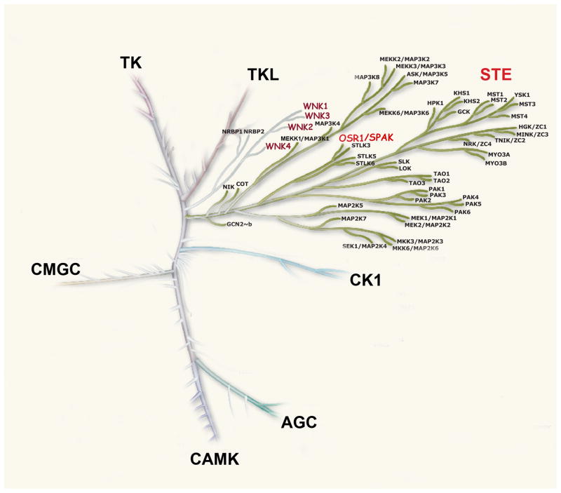
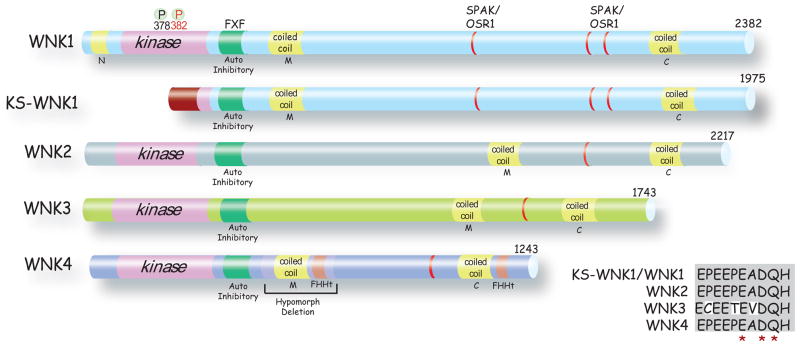
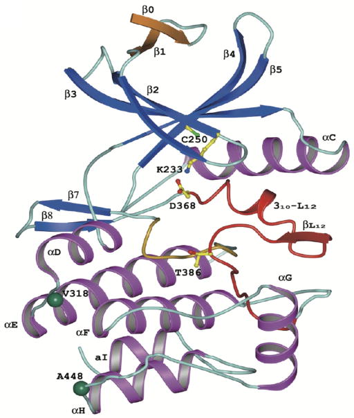
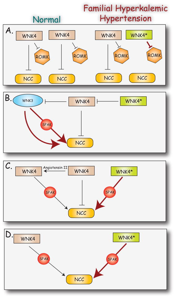
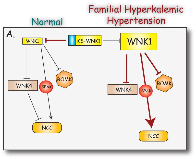
WNKs are serine/threonine kinases that comprise a unique branch of the kinome. They are so-named owing to the unusual placement of an essential catalytic lysine. WNKs have now been identified in diverse organisms. In humans and other mammals, four genes encode WNKs. WNKs are widely expressed at the message level, although data on protein expression is more limited. Soon after the WNKs were identified, mutations in genes encoding WNK1 and -4 were determined to cause the human disease familial hyperkalemic hypertension (also known as pseudohypoaldosteronism II, or Gordon's Syndrome). For this reason, a major focus of investigation has been to dissect the role of WNK kinases in renal regulation of ion transport. More recently, a different mutation in WNK1 was identified as the cause of hereditary sensory and autonomic neuropathy type II, an early-onset autosomal disease of peripheral sensory nerves. Thus the WNKs represent an important family of potential targets for the treatment of human disease, and further elucidation of their physiological actions outside of the kidney and brain is necessary. In this review, we describe the gene structure and mechanisms regulating expression and activity of the WNKs. Subsequently, we outline substrates and targets of WNKs as well as effects of WNKs on cellular physiology, both in the kidney and elsewhere. Next, consequences of these effects on integrated physiological function are outlined. Finally, we discuss the known and putative pathophysiological relevance of the WNKs.


Figures

Figure 1
Figure 1. Major families of the human kinome (adapted from (132))
WNK kinases (in red) comprise a unique branch, which is most closely related to STE kinases. Note that OSR1 (and SPAK) are members of the STE protein kinase family.
Figure 2
Figure 2. Structures of WNK Kinases
WNKs 1–4 and KS-WNK1 are shown, with the length of each, indicated. All, except KS-WNK1, contain homologous kinase domains (pink), autoinhibitory domains (green) with essential phenylalanine residues (F) separated by a single amino acid (X). All WNKs have a coiled-coil domain (yellow) near the carboxyl terminus (C) and within the middle portion of the protein (M). WNK1 also contains an amino terminal (N) coiled-coil domain. Two phosphorylated (P) serine residues that are essential for WNK1 activation are shown (378 and 382). The alternative first exon in KS-WNK1 is shown in dark red. SPAK/OSR1 binding regions are shown in red. Sites of FHHt mutations are shown in orange. The portion of the molecule deleted to generate WNK4 hypomorphic mice (167) is indicated. Inset shows an alignment of WNK kinase acidic motifs. The acidic motif is highly conserved between WNK family members. The residues mutated in WNK4 that lead to FHHt are marked *. The functional consequences of disrupting this motif in other members of the WNK family are unknown.
Figure 3
Figure 3. Kidney-specific N terminal WNK1 exon generates a kidney-specific isoform (KS-WNK1)
This isoform lacks the entire kinase domain of full length WNK1, and exon 4 is replaced by exon 4a (shown). The nucleotide and amino acid sequences of mouse and human KS-WNK1 are highly similar, (nucleotide identities indicated by *, amino acid differences indicated by red letters; amino acids indicated by single letter code). A cysteine-rich region within exon 4a is indicated by the box.
Figure 4
Figure 4. WNK1 splice variants
WNK1 displays significant variability in splicing between exons 8 and 13. The numbers above the exon boxes indicates length of each exon in nucleotides. Dashed line connecting exon indicates splicing between these exons was not determined. For variants described in O’Reilly (162), * and † indicate that these maybe identical to the variants described by Delaloy (41) since splicing 5′ of exon 10 was not determined. The renal transcripts identified O’Reilly are distinct from those identified by Shekarabi (211) since HSN-containing transcripts are neuron-specific. The functional consequences of alternative splicing are unknown. Note the additional splice variant that results in hereditary sensory and autonomic neuropathy II.
Figure 5
Figure 5. WNK3 splice variants
Alternative splicing generates two distinct variants in brain, and one in kidney. Exon 18a is present in both brain and kidney, but exon 18b is brain-specific and adds an additional 47 amino acids to the protein encoded by exon 18a. Exon 22 is skipped in kidney, resulting in an isoform lacking an 11 amino acid stretch present in brain (65). The renal isoform has been shown to stimulate activity of NCC (65). In contrast brain isoform 2 exhibits an inhibitory effect on NCC, but since NCC expression has not been detected in the brain, this is probably not physiologically relevant.
Figure 6
Figure 6. Effects of dietary potassium intake on renal WNK kinase expression
O’Reilly and colleagues (162), Lazrak and colleagues (115), and Wade and colleagues (249) all detected significant increases in the ratio of KS-WNK1/WNK1 during high potassium diet. O’Reilly and colleagues reported that high potassium intake increased renal WNK4 mRNA significantly.
Figure 7
Figure 7. Human WNK1 and KS-WNK1 promoter region structures
Delaloy and colleagues mapped multiple transcription initiation sites for WNK1 by 5′ RACE-PCR, indicated by bent arrows (41). The renal-specific promoter, PKS-WNK1, initiates expression of KS-WNK1, which lacks the WNK1 kinase domain. Horizontal lines indicate consensus transcription factor binding sites identified with the TESS program. The translation start site for P2 transcripts is indicated by *.
Figure 8
Figure 8. Human WNK4 promoter region structure
Li and colleagues analyzed the hWNK4 promoter region using TRANSFAC-TESS and Match online software (124). Bent arrows indicate transcription initiation sites; horizontal lines indicate putative transcription factor binding sites. Note the presence of two glucocorticoid responsive elements (GRE), which probably function as negative GREs to inhibit WNK4 expression (123), and the GATA-1 binding site whose acetylation upregulates WNK4 expression levels (124).
Figure 9
Figure 9. Topology of the WNK1 kinase domain (taken from (143))
The five conserved β strands are blue and α helices are magenta. The extra N-terminal β strand is shown in gold. The activation loop is shown in red and the catalytic loop in yellow. Lys-233, Cys-250, Asp-368, and Thr-386 are shown in ball-and-stick representation. Cys-250 occupies the position in subdomain II occupied by the catalytic lysine residue in other protein kinases. Instead, a lysine in subdomain I (Lys-233) provides the catalytic side chain, and results in a large cavity in the back of the ATP-binding site. The green balls represent two substrate-specificity determinant residue Val-318 and Ala-418.
Figure 10
Figure 10. Protein-protein interactions
Shows sites of interactions between WNK kinases, detected either in vitro, in cells, or in vivo. References are indicated by blue circles. A
Figure 11
Figure 11. Overview of MAPK signaling pathways, with known positions of WNK kinase action
The evidence used to generate this figure is discussed in [section III.B.]. The MAPK pathway is activated by growth factors and both environmental and intracellular stress, resulting in proliferative or stress responses through regulation of gene transcription. The ERK pathway is stimulated primarily by growth factors and tumor promoters, whereas the JNK and p38 MAPK pathways are activated by pro-inflammatory cytokines and environmental stress, including hyperosmotic stress. Members of the WNK kinase family influence MAP kinase signaling pathways as shown. EGF receptor activation stimulates WNK1 which in turn activates of ERK5. Stimulation of ERK5 by WNK1 requires MEK5 and MEKK2/3 activity, and WNK1 physically interacted MEKK2/3. WNK2 inhibits ERK1/2 activation by controlling the balance of upstream regulators of PAK1 activity, RhoA and RacI. WNK4 stimulates both ERK1/2 and p38 MAPKs in response to both hypertonic stress and stimulation with EGF. ? indicate where intermediates in the signaling pathways are unknown.
Figure 12
Figure 12. Models by which mutations in WNK4 cause FHHt
In this figure, an arrow indicates stimulation, a large arrow indicates increased stimulation, and a T shape indicates inhibition. Model A (112) shows that mutation of one of two WNK4 genes leads to a loss of inhibition of NCC and increased suppression of ROMK. Model B (278) shows that mutant WNK4 exerts a dominant-negative effect on wild type WNK4’s inhibition of WNK3 (second copy of WNK4 is omitted for clarity). The uninhibited WNK3 is shown activating NCC via both SPAK-dependent and SPAK-independent processes. Model C (198) shows that angiotensin II converts WNK4 from an inhibitory mode to a stimulatory one. Mutant WNK4 is shown acting in the stimulatory mode, even in the absence of angiotensin II. Model D (282) shows that wild type WNK4 acts to stimulate NCC. According to this model, mutant WNK4 has a simple gain-of-function effect.
Figure 13
Figure 13. Model of mechanism of WNK1-induced FHHt
In this figure, an arrow indicates stimulation, a large arrow indicates increased stimulation, and a T shape indicates inhibition. Under normal circumstances, the activity of NCC and ROMK is set by the balance between KS-WNK1 and WNK1. KS-WNK1 inhibits WNK1. WNK1 inhibits ROMK. WNK1 inhibits WNK4, which inhibits NCC. WNK1 activates SPAK, which stimulates NCC. When expression levels of WNK1 are increased, relative to KS-WNK1, WNK4 suppression is increased, SPAK activation is increased, and ROMK suppression is increased. See text for details.

References

    1. Abdallah JG, Schrier RW, Edelstein C, Jennings SD, Wyse B, Ellison DH. Loop diuretic infusion increases thiazide-sensitive Na(+)/Cl(−)-cotransporter abundance: role of aldosterone. J Am Soc Nephrol. 2001;12:1335–1341. - PubMed
    1. Abidi FE, Miano MG, Murray JC, Schwartz CE. A novel mutation in the PHF8 gene is associated with X-linked mental retardation with cleft lip/cleft palate. Clinical genetics. 2007;72:19–22. - PMC - PubMed
    1. Achard JM, Warnock DG, Disse-Nicodeme S, Fiquet-Kempf B, Corvol P, Fournier A, Jeunemaitre X. Familial hyperkalemic hypertension: phenotypic analysis in a large family with the WNK1 deletion mutation. AmJMed. 2003;114:495–498. - PubMed
    1. Adragna NC, Chen Y, Delpire E, Lauf PK, Morris M. Hypertension in K-Cl cotransporter-3 knockout mice. Adv Exp Med Biol. 2004;559:379–385. - PubMed
    1. Adragna NC, Di Fulvio M, Lauf PK. Regulation of K-Cl cotransport: from function to genes. J Membr Biol. 2004;201:109–137. - PubMed

Publication types

MeSH terms

Substances