. 2011 Feb 8;108(6):2557-62.
doi: 10.1073/pnas.1012346108. Epub 2011 Jan 19.

Action potentials drive body wall muscle contractions in Caenorhabditis elegans

Affiliations

Action potentials drive body wall muscle contractions in Caenorhabditis elegans

Shangbang Gao et al. Proc Natl Acad Sci U S A. .

Abstract

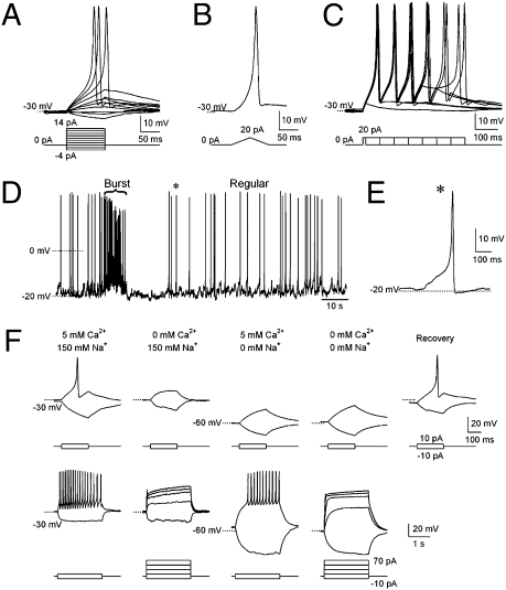
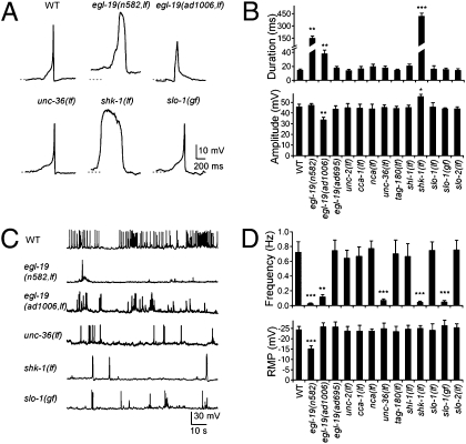
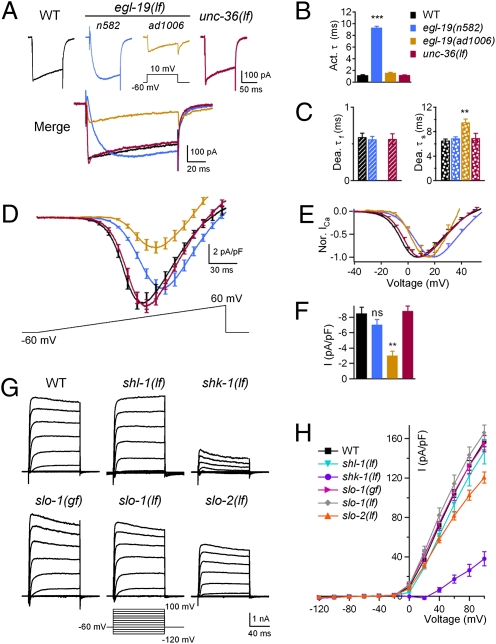
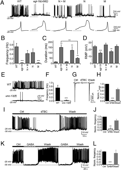
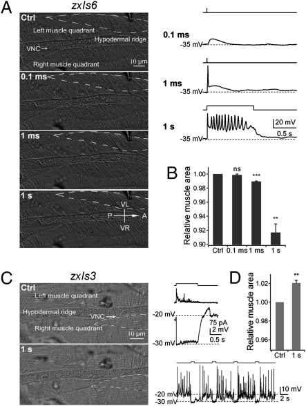
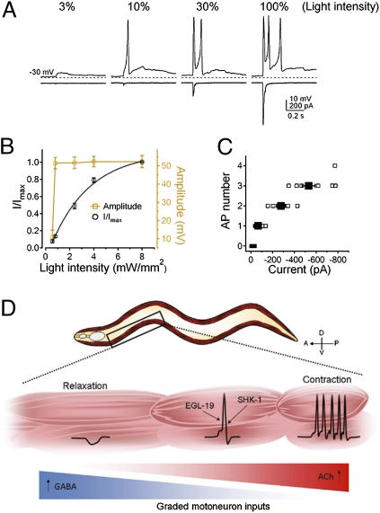
The sinusoidal locomotion exhibited by Caenorhabditis elegans predicts a tight regulation of contractions and relaxations of its body wall muscles. Vertebrate skeletal muscle contractions are driven by voltage-gated sodium channel-dependent action potentials. How coordinated motor outputs are regulated in C. elegans, which does not have voltage-gated sodium channels, remains unknown. Here, we show that C. elegans body wall muscles fire all-or-none, calcium-dependent action potentials that are driven by the L-type voltage-gated calcium and Kv1 voltage-dependent potassium channels. We further demonstrate that the excitatory and inhibitory motoneuron activities regulate the frequency of action potentials to coordinate muscle contraction and relaxation, respectively. This study provides direct evidence for the dual-modulatory model of the C. elegans motor circuit; moreover, it reveals a mode of motor control in which muscle cells integrate graded inputs of the nervous system and respond with all-or-none electrical signals.


Conflict of interest statement

The authors declare no conflict of interest.

Figures

Fig. 1.
Fig. 1.
C. elegans body wall muscles fire all-or-none action potentials. (AC) Action potentials evoked by current steps from −4 to +14 pA in 2-pA increments (A), ramp currents from 0 to +20 pA (B), and +20 pA currents from 10- to 360-ms duration in 50-ms increments (C). Dotted lines, −30 mV. (D) Spontaneous action potentials exhibit “burst” (brace) and “regular” firing modes. (E) A scaled single spontaneous action potential labeled with asterisk D. (F) Upper traces show that single action potentials were abolished in the absence of Ca2+, Na+, or both. Lower traces show that, with increased current injection time and amplitude, trains of action potentials were elicited in normal and 0 Na+ solutions, but were not in 0 Ca2+, or 0 Ca2+ and Na+ solutions.
Fig. 2.
Fig. 2.
Channels responsible for action potentials in C. elegans body wall muscles. (A) Representative single action potentials from WT and channel mutant muscles. Pronounced changes in kinetics were present in egl-19(n582,lf), egl-19(ad1006,lf), and shk-1(lf) but absent from unc-36(lf) and slo-1(gf) mutants. Dashed lines, −20 mV. (B) Quantification of the half-width and peak amplitude of action potentials in the respective channel mutants. An extension of duration was observed in egl-19(n582,lf), egl-19(ad1006,lf), and shk-1(lf). There was an increase of the peak amplitude in shk-1(lf) mutants but a decrease in egl-19(ad1006,lf) mutants. (C) Representative traces for trains of spontaneous action potentials in WT and mutant animals. (D Upper) The frequency of action potentials was decreased in egl-19(n582,lf), egl-19(ad1006,lf), unc-36(lf), shk-1(lf), and slo-1(gf) mutants. (Lower) The resting membrane potential was elevated in egl-19(n582,lf) mutants. *P < 0.05; **P < 0.01; ***P < 0.001; t test against WT. (Error bars = SEM.)
Fig. 3.
Fig. 3.
lf mutations in L-VGCC/EGL-19 and Kv1/SHK-1 affect the voltage-dependent Ca2+ and K+ currents. (A) Representative voltage-gated Ca2+ currents of WT, egl-19(n582,lf), egl-19(ad1006,lf), and unc-36(lf) mutants. (B) Ca2+ current activation time constants (activation τ) were increased in egl-19(n582,lf) mutants. (C) The fast (Dea. τf) component of Ca2+ current deactivation time constants was abolished, and the slow (Dea. τs) component was delayed in egl-19(ad1006,lf) mutants. (D) Current–voltage (IV) relationship of VGCC was affected in egl-19(n582,lf) and egl-19(ad1006,lf) but not unc-36(lf). (E) Normalized IV curve showed that the voltage dependence was shifted by approximately +10 mV in egl-19(n582,lf) and by approximately +5 mV in egl-19(ad1006,lf). (F) The peak Ca2+ current density was decreased approximately threefold in egl-19(ad1006,lf) animals but unaffected in egl-19(n582,lf) and unc-36(lf) mutants. (G and H) K+ current densities showed a significant decrease in shk-1(lf) and a slight reduction in slo-2(lf) mutants at ≥+40 mV. ns, not significant; **P < 0.01; ***P < 0.001; t test against WT. (Error bars: SEM.)
Fig. 4.
Fig. 4.
Neuronal activity modulates spontaneous action potential frequency in body wall muscles. (AD) Neuronal L-VGCC/EGL-19 modulates the frequency of action potentials, whereas muscle EGL-19 initiates action potentials. (A Upper) Single and trains of action potentials in WT, egl-19(n582), and egl-19(n582) animals expressing EGL-19 in neurons and muscles (N + M, n = 6), neurons (N, n = 3), and muscles (M, n = 5). (B) Action potential frequency was rescued in N + M and N, but not in M. (C) The duration of action potentials was rescued in N + M and partially rescued in M, but not in N. (D) The resting membrane potential was rescued in N + M and M, but not in N. (E and F) Spontaneous action potential frequency was reduced in unc-13(lf) mutants (n = 7) compared with WT (n = 10). (G) Representative traces of action potential firing in zxIs6 animals when activated by 10-ms light stimulation, treated with 0.5 mM dTBC. (H) Quantification of ACh receptor currents during and after dTBC treatment (n = 9). (I and J) Frequency of action potential firing during dTBC treatment (n = 7). (K and L) Complete and reversible inhibition of action potential firing upon 0.5 mM GABA treatment (n = 6). **P < 0.01; ***P < 0.001; t test against WT or control. (Error bars: SEM.)
Fig. 5.
Fig. 5.
Action potentials drive body wall muscle contractions. (A Left) Representative muscle morphology before and after 0.1-ms, 1-ms, and 1-s light stimulation. VNC, ventral neural cord. (Right) The membrane potential in response to light stimulation in the same muscle. (B) Normalized muscle surface cell areas outlined by dashed lines in A. (C) In zxIs3 animals, GABAergic motoneurons hyperpolarize and drive relaxation. (Left) Representative muscle morphologies before and after 1-s light stimulation. (Right) Changes in membrane potential of body muscle cells during light stimulation. Four consecutive 1-s light stimulations induced muscle hyperpolarization and completely inhibited spontaneous action potential firing (Lower Right). (D) This inhibition correlated with muscle relaxation, shown by increased cell areas (n = 4). ns, not significant; **P < 0.01; ***P < 0.001; t test against control. (Error bars: SEM.)
Fig. 6.
Fig. 6.
Action potential-driven muscle contraction and relaxation in response to graded motoneuron inputs. (A) Graded postsynaptic currents (at −30 mV) evoked all-or-none action potentials (at 0 pA). 3%, 10%, 30%, and 100% indicate the percentage of the full light stimulation. Dashed lines, −30 mV. Data were recorded from the same muscle cell every 30 s. (B) Normalized postsynaptic currents (○, n = 14) and corresponding membrane potential peak amplitude (□, n = 11) were plotted against light intensity. The normalized postsynaptic currents were fitted with a single exponential function. (C) The number of action potentials plotted against the amplitude of postsynaptic currents (n = 8). (Error bars = SEM.) (D) Graphical representation of a model: In response to graded motoneuron inputs, muscle cells fire action potentials that coordinate the contraction or relaxation along the body.

References

    1. Sulston JE, Schierenberg E, White JG, Thomson JN. The embryonic cell lineage of the nematode Caenorhabditis elegans. Dev Biol. 1983;100:64–119. - PubMed
    1. White JG, Southgate E, Thomson JN, Brenner S. The structure of the nervous system of the nematode Caenorhabditis elegans. Philos Trans R Soc Lond B Biol Sci. 1986;314:1–340. - PubMed
    1. Brenner S. The genetics of Caenorhabditis elegans. Genetics. 1974;77:71–94. - PMC - PubMed
    1. Bargmann CI. Neurobiology of the Caenorhabditis elegans genome. Science. 1998;282:2028–2033. - PubMed
    1. Davis RE, Stretton AO. Passive membrane properties of motorneurons and their role in long-distance signaling in the nematode Ascaris. J Neurosci. 1989;9:403–414. - PMC - PubMed

Publication types

MeSH terms

Substances

LinkOut - more resources