

Stromal endothelial cells directly influence cancer progression
- PMID: 21248315
- PMCID: PMC3076139
- DOI: 10.1126/scitranslmed.3001542
Stromal endothelial cells directly influence cancer progression
Abstract
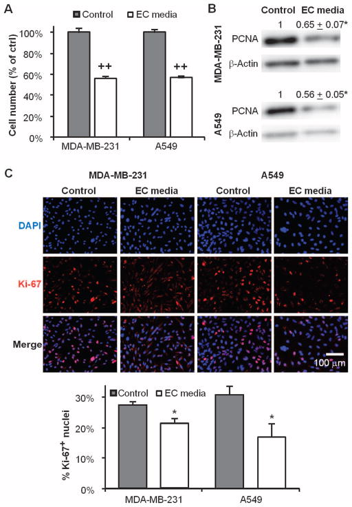
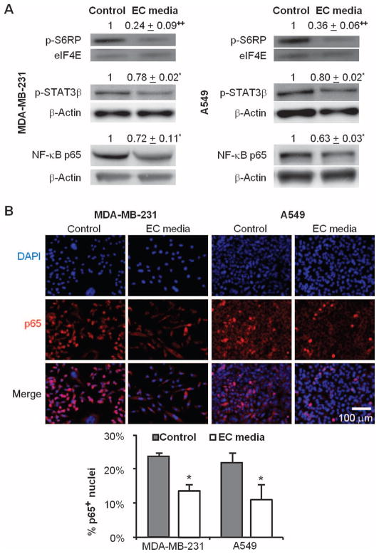
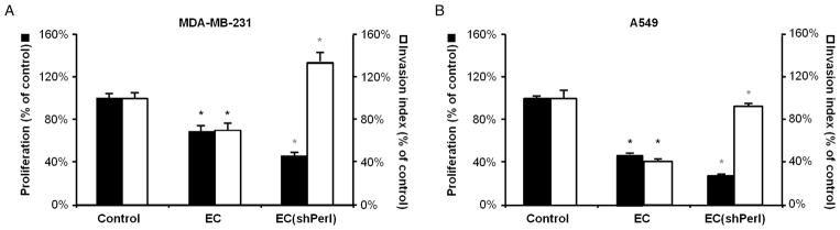
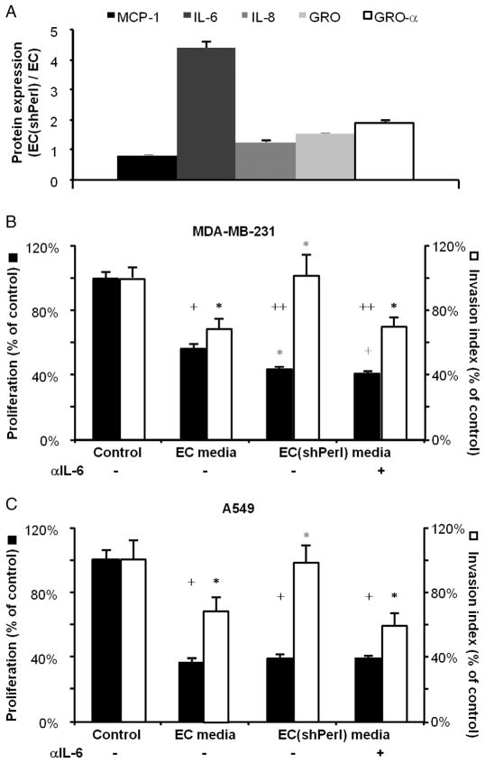
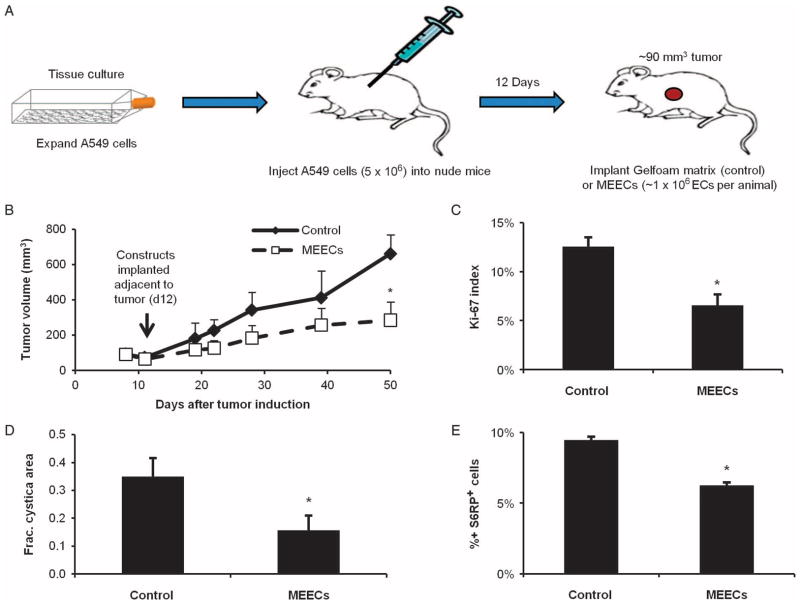
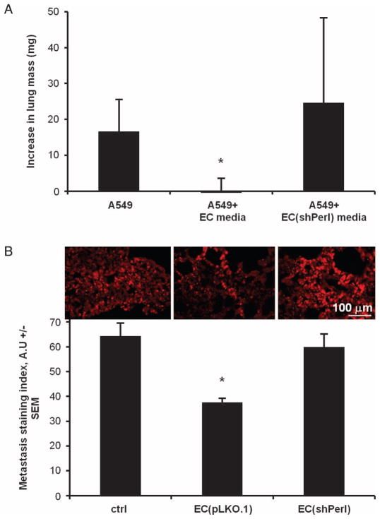
Cancer growth and metastasis are regulated in part by stromal cells such as fibroblasts and immune cells within the tumor microenvironment. Endothelial cells (ECs) are also ubiquitous within tumors because tumors are vascular, and yet, the impact of tumor-resident ECs is less well understood. Through paracrine regulation, ECs modulate a diverse spectrum of pathophysiologic processes in normal and hyperplastic tissues. We hypothesized that ECs offer similar paracrine regulatory control of cancer biology. Indeed, secretions from quiescent ECs muted the proliferative and invasive phenotype of lung and breast cancer cells in vitro and reduced cancer cell protumorigenic and proinflammatory signaling. EC perlecan silencing significantly changed this regulatory relationship, eliminating the ability of ECs to inhibit cancer cell invasiveness via increased interleukin-6 secretion. Moreover, implanting ECs embedded within porous matrices slowed adjacent xenograft tumor growth and prevented architectural degeneration, with a concomitant reduction in proliferative and tumorigenic markers. Finally, lung carcinoma cells pretreated with intact EC-conditioned media, but not media conditioned with perlecan-silenced ECs, exhibited reduced micrometastatic burden after tail vein injection. These findings add to an emerging appreciation of EC-regulatory effects that transcend their structural roles and pave the way for improved characterization and control of EC-cancer cross-talk interactions for diagnosis, prognosis, and treatment of cancer.
Figures

Similar articles
-
Dysfunctional endothelial cells directly stimulate cancer inflammation and metastasis.Int J Cancer. 2013 Sep 15;133(6):1334-44. doi: 10.1002/ijc.28146. Epub 2013 Apr 8. Int J Cancer. 2013. PMID: 23463345 Free PMC article.
-
STAT3 activation in endothelial cells is important for tumor metastasis via increased cell adhesion molecule expression.Oncogene. 2017 Sep 28;36(39):5445-5459. doi: 10.1038/onc.2017.148. Epub 2017 May 22. Oncogene. 2017. PMID: 28534515
-
Tumor‑type‑dependent effects on the angiogenic abilities of endothelial cells in an in vitro rat cell model.Oncol Rep. 2019 Jul;42(1):350-360. doi: 10.3892/or.2019.7143. Epub 2019 May 2. Oncol Rep. 2019. PMID: 31059104
-
The Multiple Roles of Exosomes in Metastasis.Cancer Genomics Proteomics. 2017 Jan 2;14(1):1-15. doi: 10.21873/cgp.20015. Cancer Genomics Proteomics. 2017. PMID: 28031234 Free PMC article. Review.
-
Involvement of Extracellular Vesicles in Vascular-Related Functions in Cancer Progression and Metastasis.Int J Mol Sci. 2019 May 26;20(10):2584. doi: 10.3390/ijms20102584. Int J Mol Sci. 2019. PMID: 31130715 Free PMC article. Review.
Cited by
-
The Differential Paracrine Role of the Endothelium in Prostate Cancer Cells.Cancers (Basel). 2022 Sep 29;14(19):4750. doi: 10.3390/cancers14194750. Cancers (Basel). 2022. PMID: 36230673 Free PMC article.
-
Chronic Obstructive Pulmonary Disease and Lung Cancer: Underlying Pathophysiology and New Therapeutic Modalities.Drugs. 2018 Nov;78(16):1717-1740. doi: 10.1007/s40265-018-1001-8. Drugs. 2018. PMID: 30392114 Review.
-
Dynamic and Cell-Infiltratable Hydrogels as Injectable Carrier of Therapeutic Cells and Drugs for Treating Challenging Bone Defects.ACS Cent Sci. 2019 Mar 27;5(3):440-450. doi: 10.1021/acscentsci.8b00764. Epub 2019 Feb 13. ACS Cent Sci. 2019. PMID: 30937371 Free PMC article.
-
The effect of substrate modulus on the growth and function of matrix-embedded endothelial cells.Biomaterials. 2013 Jan;34(3):677-84. doi: 10.1016/j.biomaterials.2012.09.079. Epub 2012 Oct 24. Biomaterials. 2013. PMID: 23102623 Free PMC article.
-
Reciprocal interactions between breast tumor and its adipose microenvironment based on a 3D adipose equivalent model.PLoS One. 2013 Jun 4;8(6):e66284. doi: 10.1371/journal.pone.0066284. Print 2013. PLoS One. 2013. PMID: 23750285 Free PMC article.
References
-
- Folkman J. Tumor angiogenesis: Therapeutic implications. N Engl J Med. 1971;285:1182–1186. - PubMed
-
- Folkman J. Angiogenesis. Annu Rev Med. 2006;57:1–18. - PubMed
-
- Jain RK. Normalization of tumor vasculature: An emerging concept in antiangiogenic therapy. Science. 2005;307:58–62. - PubMed
-
- Folkman J. Antiangiogenesis in cancer therapy—endostatin and its mechanisms of action. Exp Cell Res. 2006;312:594–607. - PubMed
Publication types
MeSH terms
Substances
Grants and funding
LinkOut - more resources
Full Text Sources
Other Literature Sources
