. 2011 Apr;36(5):1003-14.
doi: 10.1038/npp.2010.238. Epub 2011 Jan 19.

Discrete forebrain neuronal networks supporting noradrenergic regulation of sensorimotor gating

Affiliations

Discrete forebrain neuronal networks supporting noradrenergic regulation of sensorimotor gating

Karen M Alsene et al. Neuropsychopharmacology. 2011 Apr.

Abstract

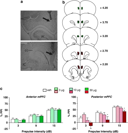
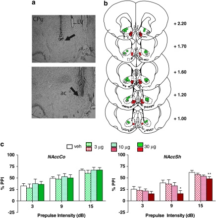
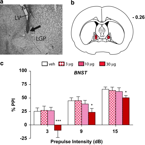
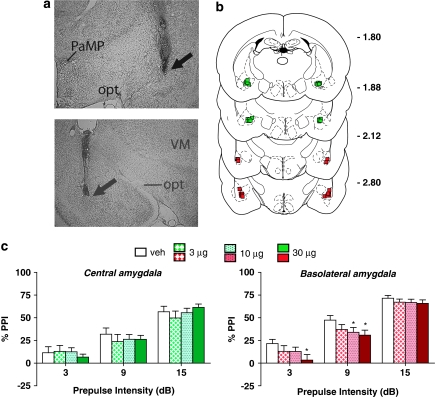
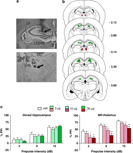
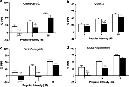
Prepulse inhibition (PPI) refers to the reduction in the startle response when a startling stimulus is preceded by a weak prestimulus, and is an endophenotype of deficient sensorimotor gating in several neuropsychiatric disorders. Emerging evidence suggests that norepinephrine (NE) regulates PPI, however, the circuitry involved is unknown. We found recently that stimulation of the locus coeruleus (LC), the primary source of NE to the forebrain, induces a PPI deficit that is a result of downstream NE release. Hence, this study sought to identify LC-innervated forebrain regions that mediate this effect. Separate groups of male Sprague-Dawley rats received a cocktail solution of the α1-NE receptor agonist phenylephrine plus the β-receptor agonist isoproterenol (equal parts of each; 0, 3, 10, and 30 μg) into subregions of the medial prefrontal cortex (mPFC), nucleus accumbens (NAcc), extended amygdala, mediodorsal thalamus (MD-thalamus), or the dorsal hippocampus (DH) before PPI testing. NE agonist infusion into the posterior mPFC, NAcc shell, bed nucleus of the stria terminalis, basolateral amygdala, and the MD-thalamus disrupted PPI, with particularly strong effects in MD-thalamus. Sites in which NE receptor stimulation did not disrupt PPI (anterior mPFC, NAcc core, central amygdala, and DH) did support PPI disruptions with the dopamine D2 receptor agonist quinpirole (0, 10 μg). This pattern reveals new pathways in the regulation of PPI, and suggests that NE transmission within distinct thalamocortical and ventral forebrain networks may subserve the sensorimotor gating deficits that are seen in disorders such as schizophrenia, Tourette syndrome, and post-traumatic stress disorder.


Figures

Figure 1
Figure 1
(a) Representative injector tip locations within the anterior (top panel) and posterior (bottom panel) medial prefrontal cortex (mPFC), indicated by the arrows. ac, anterior commissure; IC, insular cortex; cc, corpus callosum. (b) Chartings depicting the locations in which infusions of the α1+β-NE receptor agonist cocktail did (red) and did not (green) disrupt % prepulse inhibition (PPI). Distances are in mm from the bregma. (c) Effects on % PPI of the phenylephrine+isoproterenol cocktail in the anterior and posterior mPFC. Values represent means±SEM for each dose. Doses are in μg/0.5 μl. Prepulse intensity indicates decibels above the background noise level. *P<0.05, relative to VEH (vehicle) condition.
Figure 2
Figure 2
(a) Representative injector tip locations within the nucleus accumbens core (NAccCo) (top panel) and shell (NAccSh) (bottom panel), indicated by the arrows. ac, anterior commissure; CPu, caudate putamen; LV, lateral ventricle. (b) Chartings depicting the locations in which infusions of the α1+β-NE receptor agonist cocktail did (red) and did not (green) disrupt % prepulse inhibition (PPI). Distances are in mm from the bregma. (c) Effects on % PPI of the phenylephrine+isoproterenol cocktail in the NAccCo and NAccSh. Values represent means±SEM for each dose. Doses are in μg/0.5 μl. Prepulse intensity indicates decibels above the background noise level. *P<0.05, **P<0.01 relative to VEH (vehicle) condition.
Figure 3
Figure 3
(a) Representative injector tip location within the bed nucleus of the stria terminalis (BNST), indicated by the arrow. LV, lateral ventricle; LGP, lateral globus pallidus. (b) Charting depicting the locations in which infusions of the α1+β-NE receptor agonist cocktail disrupted % prepulse inhibition (PPI). Distance is in mm from the bregma. (c) Effects on % PPI of the phenylephrine+isoproterenol cocktail in the BNST. Values represent means±SEM for each dose. Doses are in μg/0.5 μl. Prepulse intensity indicates decibels above the background noise level. *P<0.05, ***P<0.01 relative to VEH (vehicle) condition.
Figure 4
Figure 4
(a) Representative injector tip locations within the central amygdala (CeA) (top panel), and the basolateral amygdala (BLA) (bottom panel), indicated by the arrows. PaMP, paraventricular hypothalamic nucleus (medial part); opt, optic tract; VM, ventromedial thalamic nucleus. (b) Chartings depicting the locations in which infusions of the α1+β-NE receptor agonist cocktail did (red) and did not (green) disrupt % prepulse inhibition (PPI). Distances are in mm from the bregma. (c) Effects on % PPI of the phenylephrine+isoproterenol cocktail in the CeA and the BLA. Values represent means±SEM for each dose. Doses are in μg/0.5 μl. Prepulse intensity indicates decibels above the background noise level. *P<0.05, relative to VEH (vehicle) condition.
Figure 5
Figure 5
(a) Representative injector tip locations within the dorsal hippocampus (DH) (top panel) and the mediodorsal (MD) thalamus (bottom panel), indicated by the arrows. LV, lateral ventricle; LDDM, laterodorsal thalamic nucleus; DG, dentate gyrus; LHb, lateral habenular nucleus. (b) Chartings depicting the locations in which infusions of the α1+β-NE receptor agonist cocktail did (red) and did not (green) disrupt % prepulse inhibition (PPI). Distances are in mm from the bregma. (c) Effects on % PPI of the phenylephrine+isoproterenol cocktail in the DH and the MD- thalamus. Values represent means±SEM for each dose. Doses are in μg/0.5 μl. Prepulse intensity indicates decibels above the background noise level. *P<0.05, **P<0.001, relative to VEH (vehicle) condition.
Figure 6
Figure 6
Effects on % prepulse inhibition (PPI) of treatment with the dopamine receptor agonist quinpirole in the (a) anterior mPFC, (b) nucleus accumbens core, (c) central amygdala, and (d) dorsal hippocampus. Values represent means±SEM for each drug condition. White bars are vehicle values and black bars are values for quinpirole (10 μg/0.5 μl). Prepulse intensity indicates decibels above the background noise level. *P<0.05, **P<0.01, ***P<0.001 relative to vehicle condition.

References

    1. Alelu-Paz R, Gimenez-Amaya JM. The mediodorsal thalamic nucleus and schizophrenia. J Psychiatry Neurosci. 2008;33:489–498. - PMC - PubMed
    1. Alsene KM, Carasso BS, Connors EE, Bakshi VP. Disruption of prepulse inhibition after stimulation of central but not peripheral alpha-1 adrenergic receptors. Neuropsychopharmacology. 2006;31:2150–2161. - PubMed
    1. Arnsten AF. Adrenergic targets for the treatment of cognitive deficits in schizophrenia. Psychopharmacology (Berl) 2004;174:25–31. - PubMed
    1. Arnsten AF, Mathew R, Ubriani R, Taylor JR, Li BM. Alpha-1 noradrenergic receptor stimulation impairs prefrontal cortical cognitive function. Biol Psychiatry. 1999;45:26–31. - PubMed
    1. Arnsten AF, Steere JC, Jentsch DJ, Li BM. Noradrenergic influences on prefrontal cortical cognitive function: opposing actions at postjunctional alpha 1 vs alpha 2-adrenergic receptors. Adv Pharmacol. 1998;42:764–767. - PubMed

Publication types

MeSH terms