

doi: 10.1038/ejhg.2010.235.
Epub 2011 Jan 19.
Craniosynostosis
Affiliations
- PMID: 21248745
- PMCID: PMC3060331
- DOI: 10.1038/ejhg.2010.235
Item in Clipboard
Craniosynostosis
Eur J Hum Genet.
2011 Apr.
Abstract
Craniosynostosis, defined as the premature fusion of the cranial sutures, presents many challenges in classification and treatment. At least 20% of cases are caused by specific single gene mutations or chromosome abnormalities. This article maps out approaches to clinical assessment of a child presenting with an unusual head shape, and illustrates how genetic analysis can contribute to diagnosis and management.
Figures
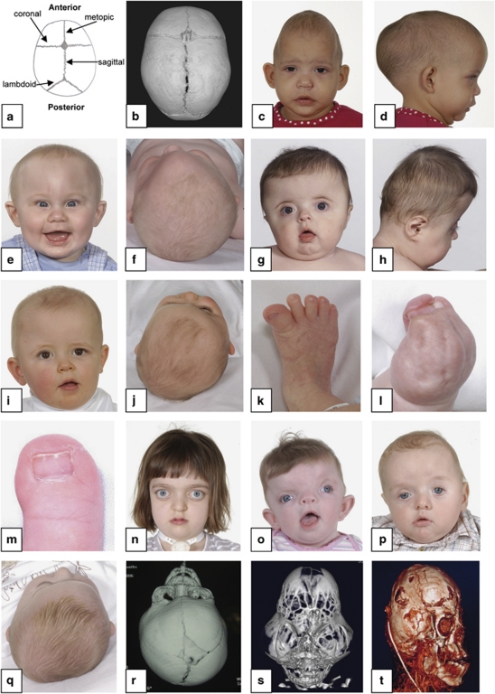
Diagnostic features of craniosynostosis. (a) Schematic diagram showing positions of the major cranial sutures. (b) CT scan (vertex view of skull) showing major sutures; anterior is at top. (c,d) Sagittal synostosis: note long, narrow head. (e,f) Metopic synostosis: note hypotelorism and triangular profile of forehead. (g,h) Bicoronal synostosis: broad, flattened head. (i,j) Right unicoronal synostosis: note flattened brow and anterior position of ear on affected side, deviation of nasal tip and prominent brow on unaffected side. (k–m), Congenital anomalies of feet or hands characteristic of Pfeiffer syndrome (k), Apert syndrome (l) and craniofrontonasal syndrome (m). (n) Crouzonoid facial appearance. (o) Severe hypertelorism, grooved nasal tip and left unicoronal synostosis in craniofrontonasal syndrome. (p) Ptosis and left unicoronal synostosis in Saethre-Chotzen syndrome. (q) Positional plagiocephaly: prominence on right anteriorly and left posteriorly, with right ear anterior and parallelogram shape to skull. (r) CT reconstruction showing left unicoronal synostosis. (s) CT reconstruction showing cloverleaf skull. (t) CT venogram showing abnormal venous drainage in multisuture syndromic craniosynostosis. See text for further details.
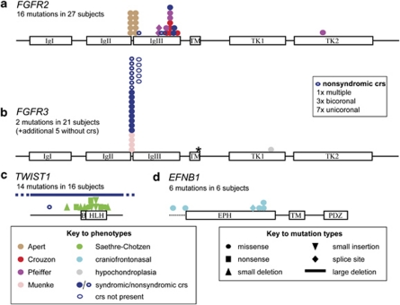
Distribution and types of mutation that commonly cause craniosynostosis. The major domains of the proteins encoded by the FGFR2 (a), FGFR3 (b), TWIST1 (c) and EFNB1 (d) genes are shown to scale, together with the position and types of mutation identified, and their associated phenotypes. Dashed line before EPH domain encoded by EFNB1 indicates 5′ untranslated region. The data, which were obtained from the Oxford cohort study, convey the relative prevalence of the most common mutations, but many rare mutations were absent in this survey. Asterisk indicates position of Ala391Glu mutation of FGFR3. crs, craniosynostosis. See text for information on protein domains.
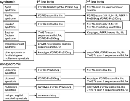
Flow diagram for molecular genetic diagnosis of craniosynostosis, showing the minimum tests recommended for each clinical presentation. In practice, the Oxford laboratory bundles sequencing of the FGFR1, FGFR2 (exons IIIa and IIIc), FGFR3 and TWIST1 genes together into a single ‘level 1' screen to simplify the workflow. If the suggested tests are negative, the diagnosis should be reviewed.
References
-
- Lajeunie E, Le Merrer M, Bonaïti-Pellie C, Marchac D, Renier D. Genetic study of nonsyndromic coronal craniosynostosis. Am J Med Genet. 1995;55:500–504. - PubMed
-
- Boulet SL, Rasmussen SA, Honein MA. A population-based study of craniosynostosis in metropolitan Atlanta, 1989–2003. Am J Med Genet. 2008;146A:984–991. - PubMed
-
- Lajeunie E, Crimmins DW, Arnaud E, Renier D. Genetic considerations in nonsyndromic midline craniosynostoses: a study of twins and their families. J Neurosurg (Pediatrics 4) 2005;103:353–356. - PubMed
-
- Haas LL. Roentgenological skull measurements and their diagnostic applications. Am J Roentgenol Radium Ther Nucl Med. 1952;67:197–209. - PubMed
Further Reading
-
- Jabs EW.TWIST1 and the Saethre-Chotzen syndromein Epstein CJ, Erickson RP, Wynshaw-Boris A (eds): Inborn Errors of Development Oxford: Oxford University Press; 2008474–481.
-
- Ornitz DM, Marie PJ. FGF signaling pathways in endochondral and intramembranous bone development and human genetic disease. Genes Dev. 2002;16:1446–1465. - PubMed
-
- Passos-Bueno MR, Sertié AL, Jehee FS, Fanganiello R. Genetics of craniosynostosis: genes, syndromes, mutations and genotype-phenotype correlations. Front Oral Biol. 2007;12:107–143. - PubMed
-
- Twigg SRF, Wilkie AOM.EFNB1 and EFNA4 in craniofrontonasal syndrome and craniosynostosisin Epstein CJ, Erickson RP, Wynshaw-Boris A (eds): Inborn Errors of Development Oxford: Oxford University Press; 20081476–1482.
-
- Webster MK, Donoghue DJ. FGFR activation in skeletal disorders: too much of a good thing. Trends Genet. 1997;13:178–182. - PubMed
Publication types
MeSH terms
Substances
Grants and funding
LinkOut - more resources
Full Text Sources
Other Literature Sources
Miscellaneous
