

Review
doi: 10.1038/nature09783.
Pervasive roles of microRNAs in cardiovascular biology
Affiliations
- PMID: 21248840
- PMCID: PMC3073349
- DOI: 10.1038/nature09783
Item in Clipboard
Review
Pervasive roles of microRNAs in cardiovascular biology
Nature.
.
Abstract
First recognized as regulators of development in worms and fruitflies, microRNAs are emerging as pivotal modulators of mammalian cardiovascular development and disease. Individual microRNAs modulate the expression of collections of messenger RNA targets that often have related functions, thereby governing complex biological processes. The wideranging functions of microRNAs in the cardiovascular system have provided new perspectives on disease mechanisms and have revealed intriguing therapeutic targets, as well as diagnostics, for a variety of cardiovascular disorders.
Figures
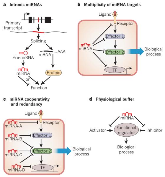
The potential modes of miRNA-based regulation of gene expression are shown. a, Intronic miRNAs are encoded within an intron of a host gene. mRNA splicing generates a protein-coding transcript and an miRNA stem–loop. Intronic miRNAs often regulate similar processes to that of the protein encoded by the host gene. AAA, polyadenylated tail of the transcript; pre-miRNA, precursor miRNA. b, A common mechanism of miRNA function involves the modest repression of several mRNAs in a common biological process by a single miRNA. This mechanism reduces the dependence on a single miRNA-mRNA interaction and increases the robustness of the gene-regulatory network. TF, transcription factor. c, Many miRNAs may cooperatively or redundantly regulate a single biological process, by individually targeting many components of that process or by synergistically repressing a crucial component of a pathway. d, miRNAs may act as a ‘buffer’ against minor perturbations in a biological pathway. This is accomplished by the targeting of factors that positively and negatively influence a particular process, thereby insulating that process from environmental fluctuations.
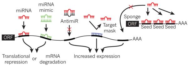
The various methods of artificially modulating miRNA expression or activity are shown. Endogenous miRNA (red) binds to complementary sequences in the 3′ UTR of a target gene, resulting in translational repression or mRNA degradation. An miRNA mimic (green) consists of an oligonucleotide duplex of the miRNA and a passenger strand. The miRNA mimic comprises the same nucleotide sequence as an endogenous miRNA, and is designed to target the same mRNAs as that miRNA. An antimiR (grey) is an oligonucleotide that is complementary to an endogenous miRNA, thereby designed to bind and inhibit its function. A target mask (blue) is an oligonucleotide designed to bind to a portion of an endogenous miRNA target without initiating mRNA degradation or translational inhibition. This strategy rescues one particular mRNA from miRNA-mediated repression. miRNA sponges consist of an open reading frame (ORF) linked to a 3′ UTR that contains several binding sites for a particular miRNA, acting as competitive inhibitors for miRNA binding.
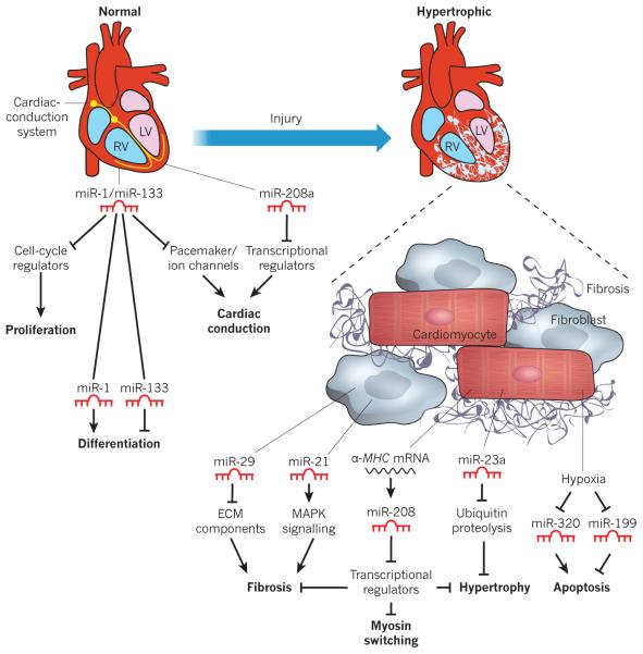
A normal and a hypertrophic heart are shown in schematic form, depicting miRNAs that contribute to normal function or pathological remodelling. The expression of selected miRNAs within the heart is shown, along with their corresponding functions. All arrows denote the normal action of each component or process. miR-1 and miR-133 are involved in the development of a normal heart (left) by regulating proliferation, differentiation and cardiac conduction. For example, proliferation is promoted by cell-cycle regulators, but miR-1 and miR-133 block these regulators, thus blocking proliferation. miR-208a also contributes to the regulation of the conduction system. After cardiac injury (right), various miRNAs contribute to pathological remodelling and the progression to heart failure. miR-29 and miR-21 block and promote cardiac fibrosis, respectively. miR-29 blocks fibrosis by inhibiting the expression of ECM components, whereas miR-21 promotes fibrosis by stimulating mitogen-activated protein kinase (MAPK) signalling. miR-208 controls myosin isoform switching, cardiac hypertrophy and fibrosis. miR-23a promotes cardiac hypertrophy by inhibiting ubiquitin proteolysis, which itself inhibits hypertrophy. Hypoxia results in the repression of miR-320 and miR-199, which promote and block apoptosis, respectively. ECM, extracellular matrix; LV, left ventricle; MHC, myosin heavy chain; RV, right ventricle.
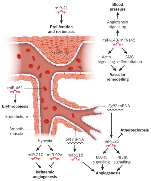
Blood vessel schematic showing the endothelial and smooth muscle layers, red blood cells and the proliferating SMCs of a neointimal lesion. The expression of select miRNAs is shown, along with their observed functional role. Hypoxia results in the activation of miR-210 and miR-92a, which promote and inhibit angiogenesis, respectively. miR-126, an endothelial-cell-enriched miRNA encoded by an intron of the Egfl7 gene, modulates atherosclerosis and angiogenesis by regulating MAPK and PI(3)K signalling. Angiogenesis is also regulated by miR-218, which is encoded by an intron of the Slit genes. miR-143 and miR-145 are expressed in SMCs and control blood pressure and vascular tone, and contribute to vascular remodelling. miR-21 is induced in SMCs after vascular injury, and promotes proliferation and neointima formation. miR-451 regulates the proliferation and differentiation of erythroid cells.
References
-
- Hill JA, Olson EN. Cardiac plasticity. N. Engl. J. Med. 2008;358:1370–1380. - PubMed
-
- Hoffman JI, Kaplan S. The incidence of congenital heart disease. J. Am. Coll. Cardiol. 2002;39:1890–1900. - PubMed
-
- Bruneau BG. The developmental genetics of congenital heart disease. Nature. 2008;451:943–948. - PubMed
-
- Latronico MV, Condorelli G. MicroRNAs and cardiac pathology. Nature Rev. Cardiol. 2009;6:419–429. - PubMed
Publication types
MeSH terms
Substances
Grants and funding
LinkOut - more resources
Full Text Sources
Other Literature Sources
