

"No performance in surgery more interesting and satisfactory": Harvey Cushing and his experience with spinal cord tumors at the Johns Hopkins Hospital
- PMID: 21250810
- PMCID: PMC4612569
- DOI: 10.3171/2010.10.SPINE10147
"No performance in surgery more interesting and satisfactory": Harvey Cushing and his experience with spinal cord tumors at the Johns Hopkins Hospital
Abstract
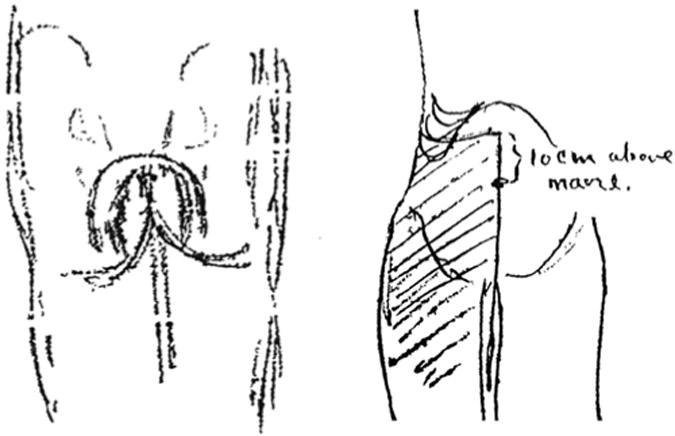
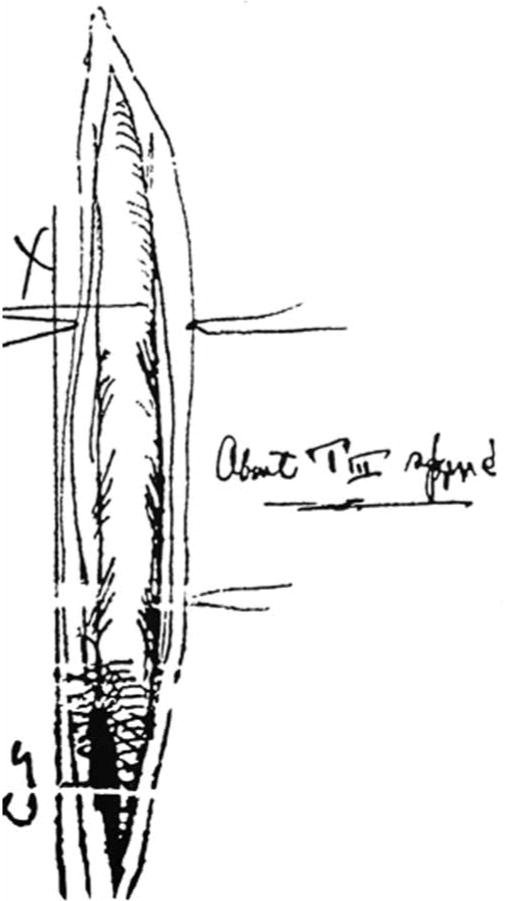
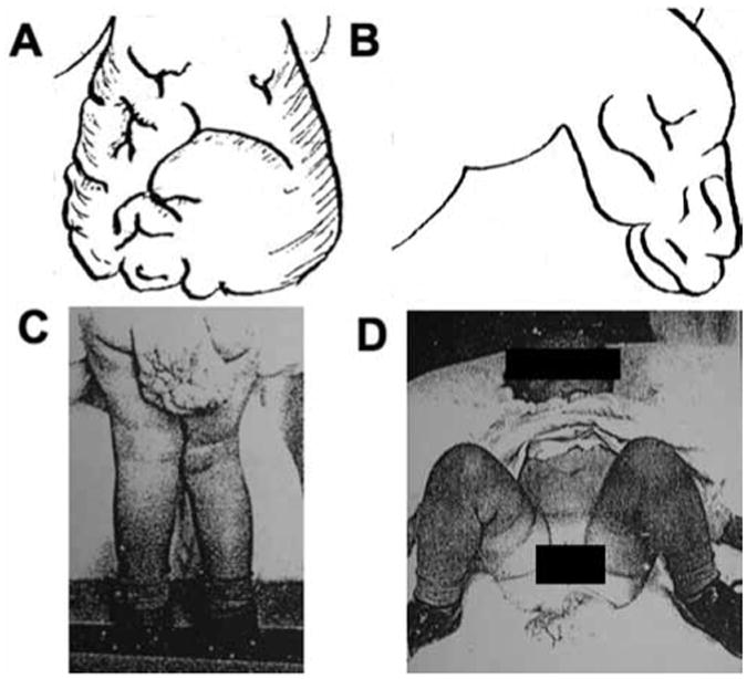
Although Harvey Cushing was a neurosurgical pioneer, his work on the spine remains largely unknown. In fact, other than his own publications, Cushing's patients with pathological lesions of the spine who were treated while he was at the Johns Hopkins Hospital, including those with spinal cord tumors, have never been previously described. The authors report on 7 patients with spinal cord tumors that Cushing treated surgically between 1898 and 1911: 2 extradural, 3 intradural extramedullary, and 2 intramedullary tumors. The authors also describe 10 patients in whom Cushing performed an "exploratory laminectomy" expecting to find a tumor, but in whom no oncological pathological entity was found. Cushing's spine surgeries were limited by challenges in making the correct diagnosis, lack of surgical precedent, and difficulty in achieving adequate intraoperative hemostasis. Other than briefly mentioning 2 of the 4 adult patients in his landmark monograph on meningiomas, these cases-both those involving tumors and those in which he performed exploratory laminectomies--have never been published before. Moreover, these cases illustrate the evolution that Harvey Cushing underwent as a spine surgeon.
Figures


Similar articles
-
Challenges in early operative approaches to intramedullary spinal cord tumors: Harvey Cushing's perspective.J Neurosurg Spine. 2015 Oct;23(4):412-8. doi: 10.3171/2014.12.SPINE13427. Epub 2015 Jun 26. J Neurosurg Spine. 2015. PMID: 26115026
-
"No clinical puzzles more interesting": Harvey Cushing and spinal trauma, the Johns Hopkins Hospital 1896-1912.Neurosurgery. 2011 Feb;68(2):420-30; discussion 430. doi: 10.1227/NEU.0b013e318201be60. Neurosurgery. 2011. PMID: 21135734 Free PMC article.
-
The development of techniques for resection of spinal cord tumors by Harvey W. Cushing.J Neurosurg Spine. 2005 Jan;2(1):92-7. doi: 10.3171/spi.2005.2.1.0092. J Neurosurg Spine. 2005. PMID: 15658135
-
Harvey Cushing's contributions to motor mapping: 1902-1912.Cortex. 2012 Jan;48(1):7-14. doi: 10.1016/j.cortex.2010.04.006. Epub 2010 Apr 29. Cortex. 2012. PMID: 20510407 Review.
-
Harvey Cushing's Wanderjahr (1900-1901).World Neurosurg. 2020 Oct;142:476-480. doi: 10.1016/j.wneu.2020.07.034. Epub 2020 Jul 19. World Neurosurg. 2020. PMID: 32698081 Free PMC article. Review.
References
-
- Bailey P. Report of two cases of tumor of the spinal cord, unaccompanied with severe pain. J Nerv Ment Dis. 1896;21:171–178.
-
- Barksdale EM, Jr, Obokhare I. Teratomas in infants and children. Curr Opin Pediatr. 2009;21:344–349. - PubMed
-
- Black PM. Harvey Cushing at the Peter Bent Brigham Hospital. Neurosurgery. 1999;45:990–1001. - PubMed
-
- Burchiel K. Cushing and Bovie: lessons learned. J Neurosurg. 2005;102:599–600. - PubMed
-
- Caponotto Removal of an intradural spinal tumor. Ann Surg. 1893;18:69.
Publication types
MeSH terms
Personal name as subject
- Actions
Grants and funding
LinkOut - more resources
Full Text Sources
