. 2011 Jul;13(6):712-21.
doi: 10.3109/14653249.2010.548380. Epub 2011 Jan 20.

Human umbilical cord blood plasma can replace fetal bovine serum for in vitro expansion of functional human endothelial colony-forming cells

Affiliations

Human umbilical cord blood plasma can replace fetal bovine serum for in vitro expansion of functional human endothelial colony-forming cells

Lan Huang et al. Cytotherapy. 2011 Jul.

Abstract

Background aims: A hierarchy of endothelial colony-forming cells (ECFC) with different levels of proliferative potential has been identified in human circulating blood and blood vessels. ECFC has recently become an attractive target for new vascular regenerative therapies; however, in vitro expansion of ECFC typically depends on the presence of fetal bovine serum (FBS) or fetal calf serum (FCS) in the culture medium, which is not appropriate for its therapeutic application.

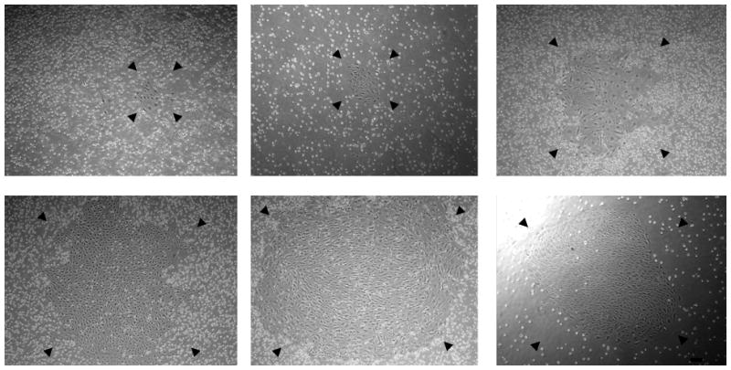
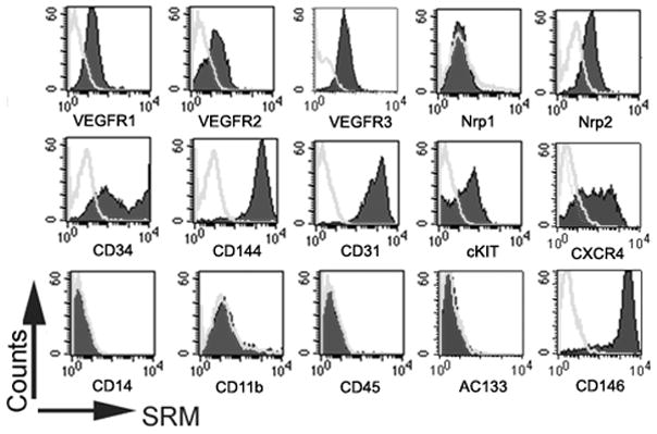
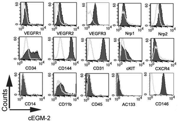
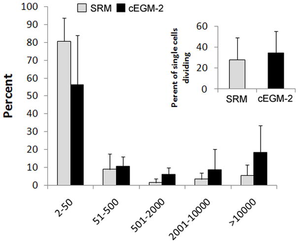
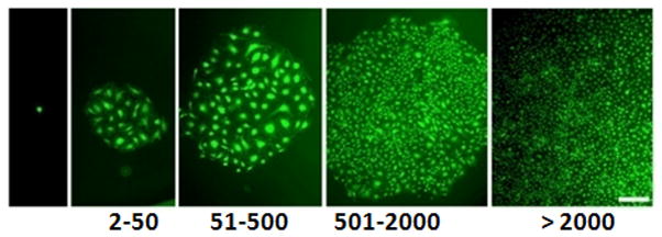
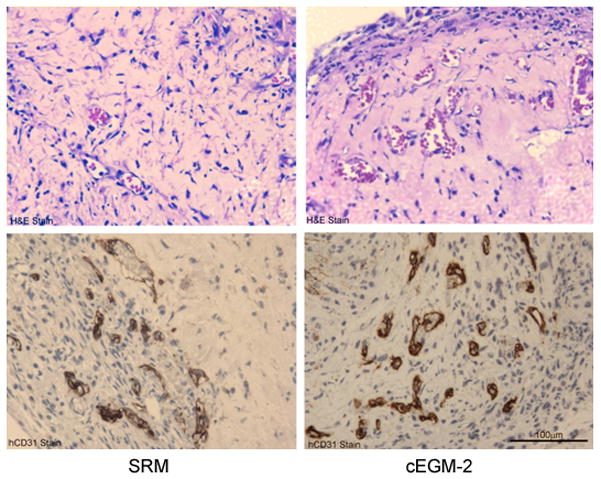
Methods: To identify optimal conditions for in vitro expansion of ECFC, the effects of human endothelial serum-free medium (SFM) supplemented with six pro-angiogenic cytokines and human umbilical cord blood plasma (HCP) were investigated. The in vitro morphology, proliferation, surface antigen expression and in vivo vessel-forming ability were utilized for examining the effects of medium on ECFC.

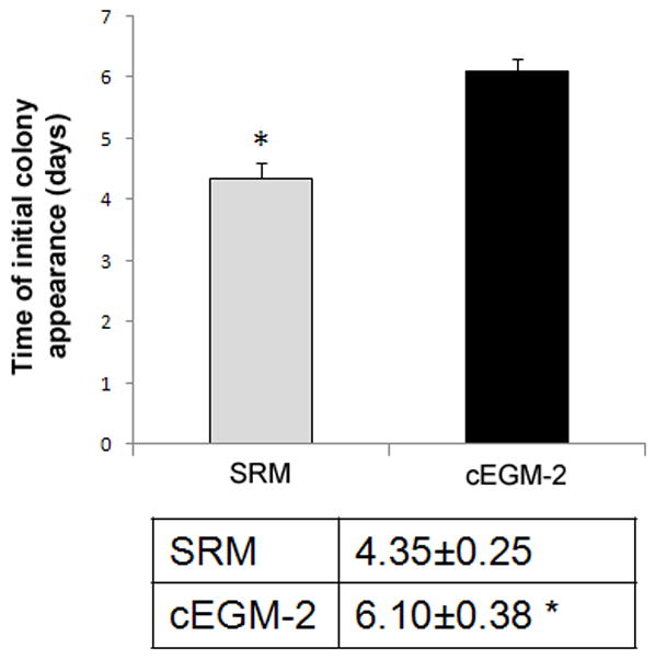
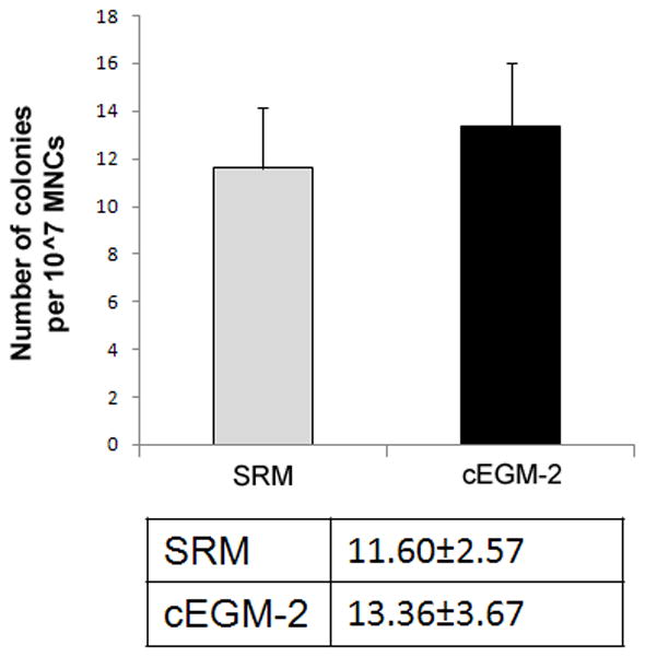
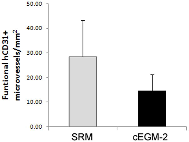
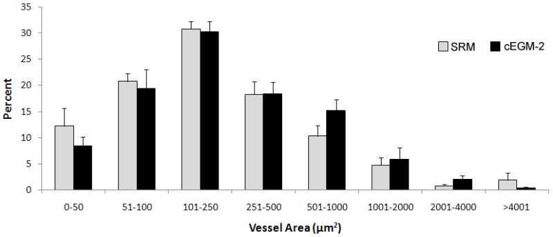
Results: This novel formulation of endothelial cell culture medium allows us, for the first time, to isolate and expand human ECFC efficiently in vitro with a low concentration of HCP (1.5%) and without bovine serum additives. In this serum-reduced medium (SRM), human ECFC colony yields remained quantitatively similar to those cultured in a high concentration (10%) of bovine serum-supplemented medium. SRM-cultured ECFC displayed a robust clonal proliferative ability in vitro and human vessel-forming capacity in vivo.

Conclusions: The present study provides a novel method for the expansion of human ECFC in vitro and will help to advance approaches for using the cells in human therapeutic trials.


Conflict of interest statement

DISCLOSURE OF INTEREST

Dr. Mervin C. Yoder is a co-founder and receives consulting fees from EndGenitor Technologies, Inc.

Figures

Figure 1
Figure 1. Isolation of human cord blood ECFC-derived EC colonies from UCB MNCs by using SRM
(A) Time of initial ECFC derived EC colonies emerged from MNCs after culture initiation in SRM and cEGM-2. Results represent the mean number of days before initial EC appearance ± SEM (Blood sample number N = 23, *P < 0.001). (B) Number of ECFC-derived EC colonies outgrown per 107 MNCs after 10 days of culture initiation in SRM and cEGM-2. Results represent the average number of EC colonies ± SEM (Blood sample number N = 23). (C) Representative photomicrographs of individual human ECFC-derived EC colonies from UCB in SRM. Scale bar represents 100 μm.
Figure 2
Figure 2. Phenotypic analysis of human cord blood ECFC-derived ECs cultured in SRM
Immunophenotyping of EC from the cultured monolayer derived from human cord blood ECFC in SRM (A) and cEGM-2 (B) by fluorescence cytometry. Similar to cells grown in cEGM-2, the ECs cultured in SRM expressed CD31, CD34, CD144, CD146, VEGFR1, VEGFR2, VEGFR3, and Nrp2 but not CD45, CD14, CD11b or AC133. Moreover, the expression of cKIT and CXCR4 was detectable in ECs grown in SRM, but not in cEGM-2.
Figure 3
Figure 3. Quantitation of the clonogenic and proliferative potential of single ECs derived from human cord blood cultured in SRM
(A) The distribution of colony sizes, where colonies were derived from single ECs grown in individual wells after 14 days of culture. The complete hierarchy of ECFCs was present in ECs cultured in SRM; that is similar to those grown in cEGM-2. Inset chart is the percentage of single ECs dividing at least once after growing 14 days in culture. No statistical difference in this frequency was observed between cells grown in SRM and those in cEGM-2. (N = 5) (B) Representive photomicrographs of EC colonies with varied sizes derived from single ECs. Scale bar represents 100μm.
Figure 4
Figure 4. Human cord blood ECFC-derived ECs cultured in SRM demonstrate the potential to form functional microvessels in immunodeficient mice
(A) H&E staining indicates microvessel formation in collagen-fibronectin gel after 14 days of implantation in NOD/SCID mice. Anti-human CD31 staining further confirms the human origin of these vessels. Scale bar represents 100 μm. (B) The number of vessels formed by human cord blood derived ECFCs (from a pool of 5 different blood samples) and perfused with murine red blood cells per mm2 in the gel after 14 days of implantation. (n=6) (C) The size distribution of the microvessels formed by human cord blood ECFCs. These data indicate that there is no difference in the vessel-forming abilities of ECs cultured in SRM versus those in cEGM-2.

References

    1. Asahara T, et al. Isolation of putative progenitor endothelial cells for angiogenesis. Science. 1997;275(5302):964–7. - PubMed
    1. Rafii S, Lyden D. Therapeutic stem and progenitor cell transplantation for organ vascularization and regeneration. Nat Med. 2003;9(6):702–12. - PubMed
    1. Hirschi KK, Ingram DA, Yoder MC. Assessing identity, phenotype, and fate of endothelial progenitor cells. Arterioscler Thromb Vasc Biol. 2008;28(9):1584–95. - PMC - PubMed
    1. Khakoo AY, Finkel T. Endothelial progenitor cells. Annu Rev Med. 2005;56:79–101. - PubMed
    1. Kovacic JC, et al. Endothelial progenitor cells, angioblasts, and angiogenesis--old terms reconsidered from a current perspective. Trends Cardiovasc Med. 2008;18(2):45–51. - PubMed

Publication types