Review
. 2011 Jan;16(1):85-95.
doi: 10.1111/j.1542-474X.2010.00399.x.

EGC diagnosis of paroxysmal supraventricular tachycardias in patients without preexcitation

Affiliations
Review

EGC diagnosis of paroxysmal supraventricular tachycardias in patients without preexcitation

Esteban González-Torrecilla et al. Ann Noninvasive Electrocardiol. 2011 Jan.

Abstract

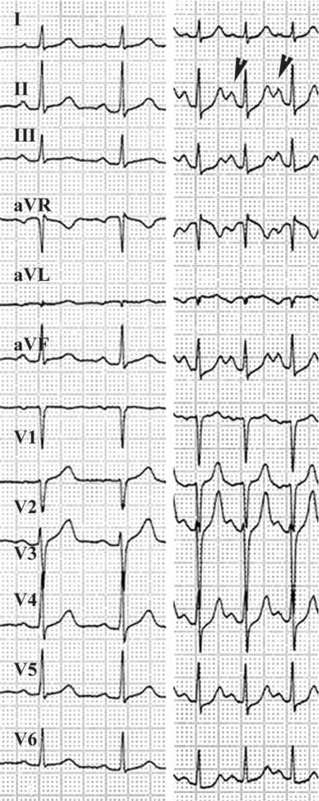
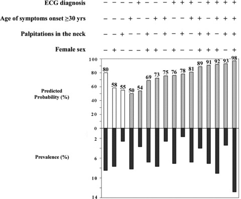
This review is aimed at discussing the diagnostic value of the different electrocardiographic criteria so far described in the differential diagnosis of the major forms of paroxysmal supraventricular tachycardias (PSVTs). The predictive value of different combinations of these independent electrocardiographic (ECG) signs in distinguishing atrioventricular reentrant tachycardias (AVRTs) through a concealed accessory pathway (AP) versus atrioventricular nodal reentrant tachycardias (AVNRTs) are discussed in detail. In addition, the adjunctive diagnostic value of simple, bedside clinical variables and their combinations to the ECG interpretation in differentiating both tachycardia mechanisms is also reviewed.


Figures

Figure 1
Figure 1
Identifying possible retrograde P waves. Careful comparison of repolarization morphologies permit probable retrograde P waves identification (arrows) and their disappearance (asterisks) with spontaneous tachycardia termination. (A) AVRT through concealed left free‐wall AP; (B) spontaneous block in retrograde fast AV nodal pathway in a patient with common AVNRT (disappearance of pseudo‐r′ wave in V1); (C) spontaneous block in retrograde slow AVN pathway in uncommon AVNRT.
Figure 2
Figure 2
Automatic focal junctional tachycardia in a 2‐year‐old infant showing clear atrioventricular dissociation in an otherwise narrow QRS complex tachycardia. Irregular firing of the automatic parahissian focus explained the irregularity of R–R intervals.
Figure 3
Figure 3
Subtle morphology changes in sinus P waves are observed during sinus tachycardia (right). At fast rates, the P wave in the inferior leads is taller because the location of the sinus pacemaker is higher in this patient with a postural orthostatic tachycardia syndrome (POTS).
Figure 4
Figure 4
Three examples of QRS alternans in AVNRT patients. Top panel: QRS alternans associated with alternative R–R intervals. Middle and bottom panels: transient QRS alternans with spontaneous disappearance (end of arrows) without discernible changes in R–R intervals.
Figure 5
Figure 5
Prevalences and univariate comparisons of major ECG criteria for AVNRT and AVRT through a concealed AP (figures are percentages of recordings with positive ECG criterion). Data from 500 consecutive ECG recordings. P < 0.01 for every criterion comparison. Abbreviations as in Figure 1.
Figure 6
Figure 6
(A) Diagnostic yield of subjective ECG interpretation in the differential diagnosis of AVNRT versus AVRT from 420 consecutive ECG tracings in patients without preexcitation during sinus rhythm who underwent invasive diagnosis. Sensitivity, specificity, and predictive values for a subjective ECG diagnosis of AVNRT (vs AVRT) are shown. (B) Corresponding values from a logistic regression model including bedside clinical variables to predict a correct AVNRT diagnosis (vs AVRT). Figures are percentages. See text for discussion.
Figure 7
Figure 7
Predicted probabilities from a logistic regression model for the diagnosis of AVNRT (gray bars) or AVRT (white bars) depending on every combination of selected ECG criteria (present: +; absent: −). The corresponding prevalences of every combination of ECG criteria in our study group are shown in dark gray bars. The predictive value of QRS alternans as an isolated ECG finding was considered as indeterminate. Figures are percentages. (From González‐Torrecilla E et al. Independent predictive accuracy of classical electrocardiographic criteria in the diagnosis of paroxysmal atrioventricular reciprocating tachycardias in patients without preexcitation. Europace 2008;10:624–8. With permission).
Figure 8
Figure 8
Predicted probabilities for the diagnosis of AVNRT (gray bars) or AVRT (white bars) depending on every combination of selected clinical covariates (present: +; absent: −). The corresponding prevalences of every combination of clinical criteria are shown in dark gray bars. For reasons of simplicity, age at symptoms onset was dichotomized using the selected cut‐off value of ≥30 years. Figures are percentages.
Figure 9
Figure 9
Predicted probabilities for the diagnosis of AVNRT (gray bars) or AVRT (white bars) depending on every combination of selected clinical covariates when ECG presumptive diagnosis is included in the logistic model. ECG is + or – indicating a diagnosis of AVNRT or AVRT, respectively.
Figure 10
Figure 10
Twelve‐lead ECGs from five patients with atypical forms of AVNRT misclassified as AVRT after subjective ECG interpretation of identifiable P waves (arrowheads).
Figure 11
Figure 11
Gender differences and prevalences of neck palpitations during tachycardia episodes in patients with common AVNRT, atypical AVNRT (slow–slow and uncommon AVNRT forms) and AVRT. The percentage of atypical AVNRT patients referring palpitations in the neck during tachycardia episodes was quite similar to those with AVRT (19%). Age at the onset of symptoms was higher in atypical AVNRT than in AVRT patients (41 ± 19 vs 25.5 ± 16 years, respectively). Figures are percentages.

References

    1. Porter MJ, Morton JB, Denman R, et al Influence of age and gender on the mechanism of supraventricular tachycardia. Heart Rhythm 2004;1:397–398. - PubMed
    1. Kalbfleisch SJ, El‐Atassi R, Calkins H, et al Differentiation of paroxysmal narrow QRS complex tachycardias using the 12‐lead electrocardiogram. J Am Coll Cardiol 1993;21:85–89. - PubMed
    1. Riva SI, Della Bella P, Fassini G, et al Value of analysis of ST segment changes during tachycardia in determining type of narrow QRS complex tachycardias. J Am Coll Cardiol 1996;27:1480–1485. - PubMed
    1. Erdinler I, Okmen E, Oguz E, et al Differentiation of narrow QRS complex tachycardia types using the 12‐lead electrocardiogram. Ann Noninvasive Electrocardiol 2002;7:120–126. - PMC - PubMed
    1. González‐Torrecilla E, Almendral J, Arenal A, et al Independent predictive accuracy of classical electrocardiographic criteria in the diagnosis of paroxysmal atrioventricular reciprocating tachycardias in patients without pre‐excitation. Europace 2008;10:624–628. - PubMed

MeSH terms