. 2011 Jan 20:12:50.
doi: 10.1186/1471-2164-12-50.

False negative rates in Drosophila cell-based RNAi screens: a case study

Affiliations

False negative rates in Drosophila cell-based RNAi screens: a case study

Matthew Booker et al. BMC Genomics. .

Abstract

Background: High-throughput screening using RNAi is a powerful gene discovery method but is often complicated by false positive and false negative results. Whereas false positive results associated with RNAi reagents has been a matter of extensive study, the issue of false negatives has received less attention.

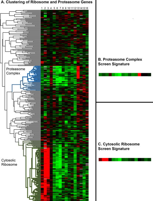
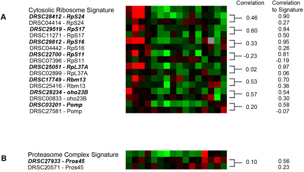
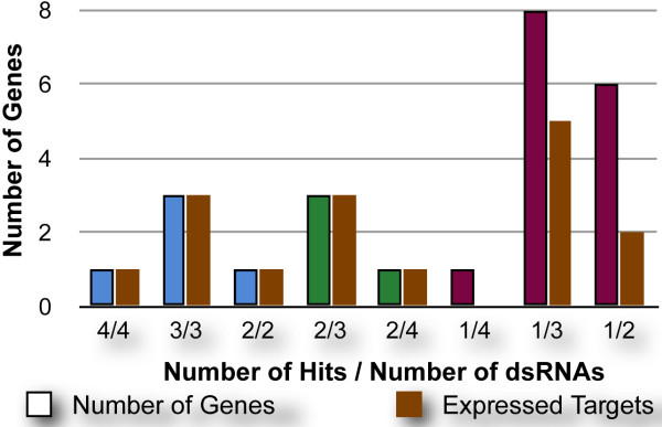
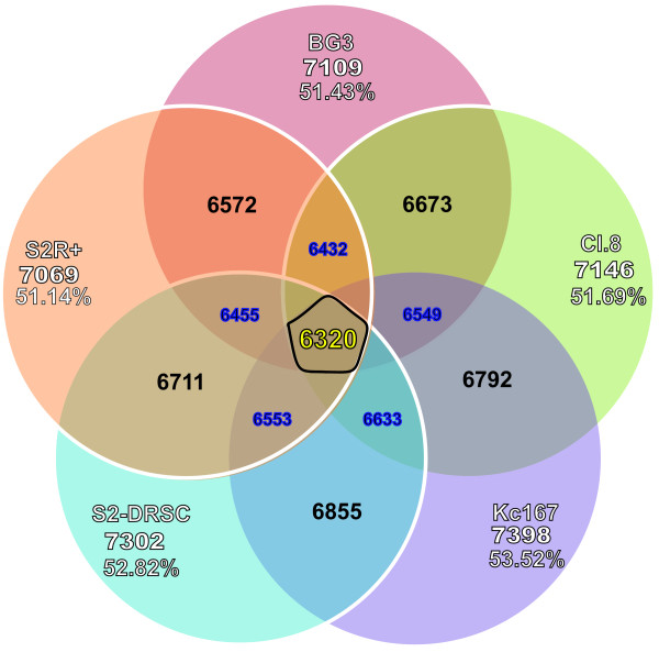
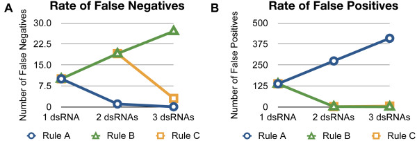
Results: We performed a meta-analysis of several genome-wide, cell-based Drosophila RNAi screens, together with a more focused RNAi screen, and conclude that the rate of false negative results is at least 8%. Further, we demonstrate how knowledge of the cell transcriptome can be used to resolve ambiguous results and how the number of false negative results can be reduced by using multiple, independently-tested RNAi reagents per gene.

Conclusions: RNAi reagents that target the same gene do not always yield consistent results due to false positives and weak or ineffective reagents. False positive results can be partially minimized by filtering with transcriptome data. RNAi libraries with multiple reagents per gene also reduce false positive and false negative outcomes when inconsistent results are disambiguated carefully.


Figures

Figure 1
Figure 1
Identification of Proteosome and Ribosome signatures in RNAi screens. All dsRNAs included in the dendrogram target 243 proteasome and ribosome-related genes. Red indicates an increase in signal and green indicates a decrease in signal. (A) Results of clustering RNAi phenotypes in 16 screens of dsRNAs targeting ribosome and proteasome genes as defined by GO terms (see Materials and Methods). The proteasome complex and cytosolic ribosome clusters are highlighted in blue and green, respectively. The simple majority of dsRNAs outside these two clusters target mitochondrial ribosome components. (B) Consensus screen signature of the proteasome complex cluster. Each small square represents the mean Z-score of the dsRNAs in the proteasome complex cluster across a single screen. (C) Consensus screen signature of the cytosolic ribosome cluster. The 16 screens are as follows from the left to the right: 1. Hormone receptor screen, plate-reader (unpublished), 2. Oncogenesis screen, plate-reader (unpublished), 3. Protein degradation screen, plate-reader (unpublished), 4. RNA processing screen, plate-reader (unpublished), 5. Mitochondrial calcium ion and proton antiporter screen, plate-reader [37], 6. Toxicity screen, plate-reader (unpublished), 7. Dengue virus host factors screen, image-based [38], 8. Ion homeostasis screen, plate-reader (unpublished). 9. Pathogen infection screen, image-based (unpublished), 10. Signaling pathway screen, plate-reader (unpublished), 11. Ion transport screen, plate-reader (unpublished), 12. Cytoskeleton regulation screen, image-based (unpublished), 13. Chromatin regulation screen, image-based (unpublished), 14. Francisella tularensis infection screen, plate-reader [39], 15. mRNA processing screen, plate-reader (unpublished), 16. Protein secretion screen, plate-reader (unpublished).
Figure 2
Figure 2
Estimation of the rate of false negatives for the Ribosome (A) and Proteasome (B) set. Red indicates an increase in signal and green indicates a decrease in signal. (A) The cytosolic ribosome screen signature is compared to the screen signatures in those cases where one dsRNA is part of the cytosolic ribosome cluster and the other is not. dsRNAs with a screen signature similar to the consensus cytosolic ribosome signature are italicized. Pearson's correlation is shown between dsRNAs that target the same gene as well as the correlation between each dsRNA and the consensus signature. (B) Similar comparison for the proteasome complex screen signature.
Figure 3
Figure 3
Results of the JAK/STAT signaling screen. The number of genes binned by the number of dsRNAs that scored out of the number of dsRNAs screened is shown. These are color-coded further: Blue for category 1 in which all dsRNAs scored, Green for category 2 in which at least two dsRNAs scored and maroon for category 3 in which only one dsRNA scored. The beige column to the right indicates the number of genes in each binned category that are expressed in S2R+ cells.
Figure 4
Figure 4
Transformed expression levels of core components of JAK-STAT signaling pathway. Genes expressed at low and high levels are displayed in gradations of black and red, correspondingly.
Figure 5
Figure 5
Number of genes expressed in different cell lines at FPKM levels greater than one. The cell lines included in the analysis are Kc167, Clone8, S2, BG3, and S2R+. 6,320 genes are expressed in all five cell lines.
Figure 6
Figure 6
Number of False Negatives and False Positives under hypothetical screening scenarios. We assume a false positive rate of 1% and a false negative rate of 10%, a scenario of 100 "true hits" in the library, and a library targeting 13,735 protein-encoding genes. (A) The predicted number of false negatives with 1, 2, or 3 dsRNAs per gene under 3 different rules for interpreting ambiguous cases. (B) The predicted number of false positives with 1, 2, or 3 dsRNAs per gene under 3 different rules for interpreting ambiguous cases.

References

    1. Echeverri CJ, Perrimon N. High-throughput RNAi screening in cultured cells: a user's guide. Nat Rev Genet. 2006;7:373–84. doi: 10.1038/nrg1836. - DOI - PubMed
    1. Echeverri CJ, Beachy PA, Baum B, Boutros M, Buchholz F, Chanda SK, Downward J, Ellenberg J, Fraser AG, Hacohen N, Hahn WC, Jackson AL, Kiger A, Linsley PS, Lum L, Ma Y, Mathey-Prévôt B, Root DE, Sabatini DM, Taipale J, Perrimon N, Bernards R. Minimizing the risk of reporting false positives in large-scale RNAi screens. Nat Methods. 2006;3:777–9. doi: 10.1038/nmeth1006-777. - DOI - PubMed
    1. Mohr S, Bakal C, Perrimon N. Genomic screening with RNAi: Results and challenges. Annu Rev Biochem. 2010;79:37–64. doi: 10.1146/annurev-biochem-060408-092949. - DOI - PMC - PubMed
    1. Zhang XD, Heyse JF. Determination of sample size in genome-scale RNAi screens. Bioinformatics. 2009;25:841–4. doi: 10.1093/bioinformatics/btp082. - DOI - PubMed
    1. Zhang XD. A new method with flexible and balanced control of false negatives and false positives for hit selection in RNA interference high-throughput screening assays. J Biomol Screen. 2007;12:645–55. doi: 10.1177/1087057107300645. - DOI - PubMed

Publication types

Substances