Review
. 2011 Jan 20:9:13.
doi: 10.1186/1479-5876-9-13.

Etiopathology of chronic tubular, glomerular and renovascular nephropathies: clinical implications

Affiliations
Review

Etiopathology of chronic tubular, glomerular and renovascular nephropathies: clinical implications

José M López-Novoa et al. J Transl Med. .

Abstract

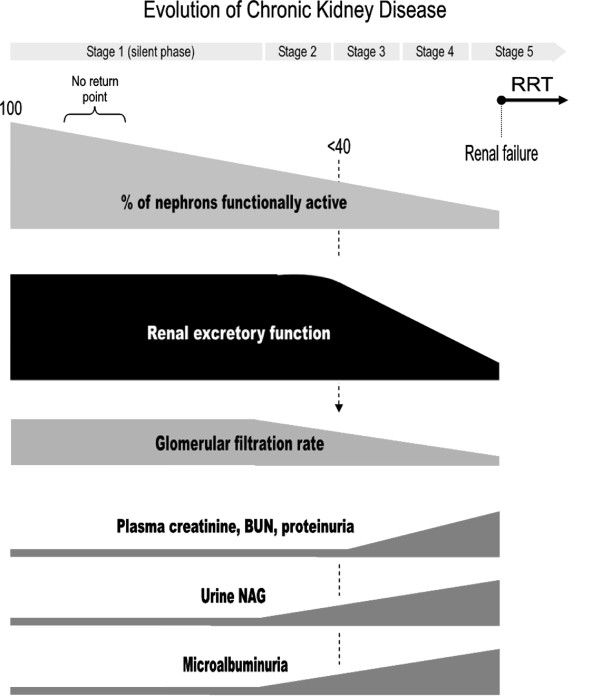
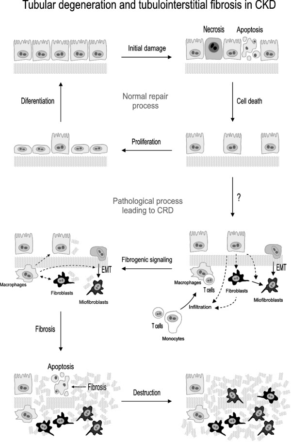
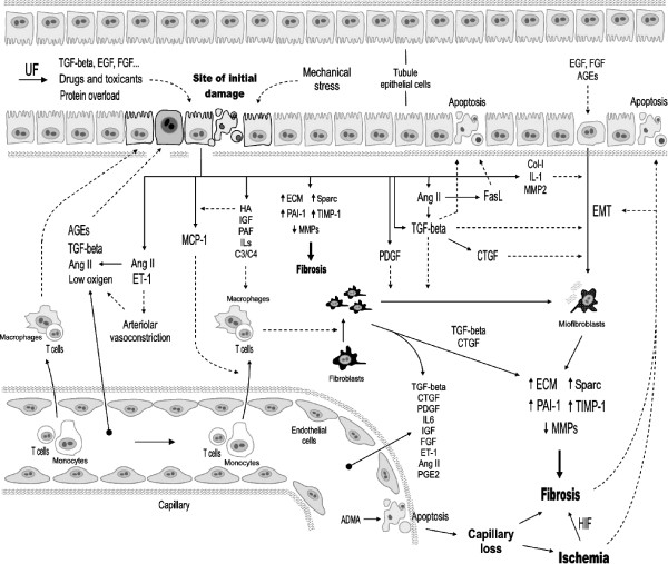
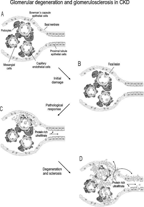
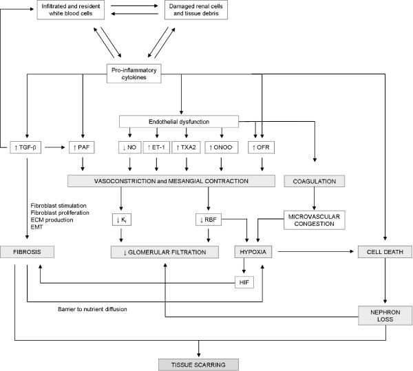
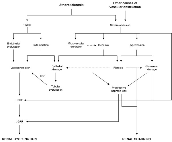
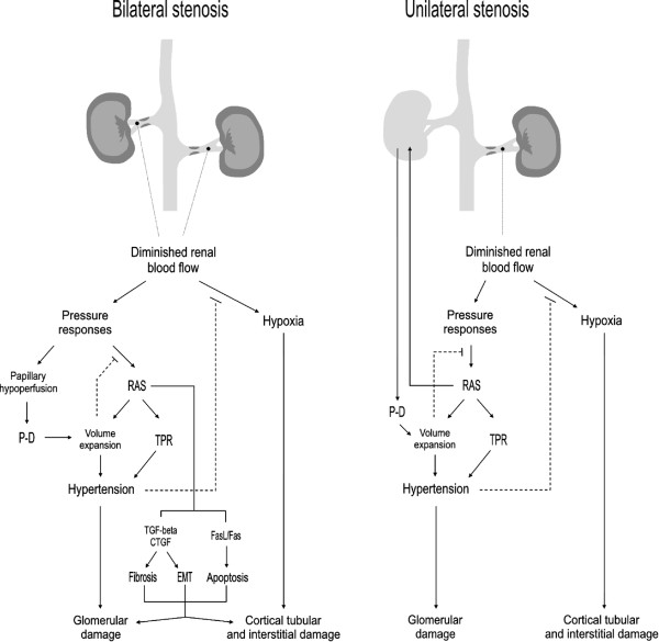
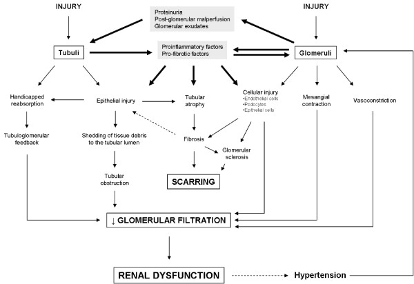
Chronic kidney disease (CKD) comprises a group of pathologies in which the renal excretory function is chronically compromised. Most, but not all, forms of CKD are progressive and irreversible, pathological syndromes that start silently (i.e. no functional alterations are evident), continue through renal dysfunction and ends up in renal failure. At this point, kidney transplant or dialysis (renal replacement therapy, RRT) becomes necessary to prevent death derived from the inability of the kidneys to cleanse the blood and achieve hydroelectrolytic balance. Worldwide, nearly 1.5 million people need RRT, and the incidence of CKD has increased significantly over the last decades. Diabetes and hypertension are among the leading causes of end stage renal disease, although autoimmunity, renal atherosclerosis, certain infections, drugs and toxins, obstruction of the urinary tract, genetic alterations, and other insults may initiate the disease by damaging the glomerular, tubular, vascular or interstitial compartments of the kidneys. In all cases, CKD eventually compromises all these structures and gives rise to a similar phenotype regardless of etiology. This review describes with an integrative approach the pathophysiological process of tubulointerstitial, glomerular and renovascular diseases, and makes emphasis on the key cellular and molecular events involved. It further analyses the key mechanisms leading to a merging phenotype and pathophysiological scenario as etiologically distinct diseases progress. Finally clinical implications and future experimental and therapeutic perspectives are discussed.


Figures

Figure 1
Figure 1
Graphic representation of the evolution of key pathological events, such as percentage of nephrons functionally active, overall renal excretory function and glomerular filtration rate, and plasma and urine markers associated with time course of chronic kidney disease. The figure shows the relative priority of appearance of these elements with repect to one another as it occurs in most cases of chronic kidney diseases. Their appearance, however, may vary from this general prototype in specific diseases or in determined cases. In the same way, the slope of increase or decline may also vary. RRT: renal replacement therapy, BUN: blood urea nitrogen, NAG: N-acetyl-β-D-glucosaminidase.
Figure 2
Figure 2
Schematic depiction of the pathological process of tubular degeneration and tubulointerstitial fibrosis characteristic of tubulointerstitial diseases, and also of later stages of glomerular and renovascular diseases leading to chronic kidney disease (adapted from references [87]and [291]). EMT, epithelial to mesenchymal transition.
Figure 3
Figure 3
Extracellular mediators and effectors of tubulointerstitial pathological events in chronic kidney disease. ADMA: asymmetric dimethylarginine. HA, hyaluronic acid. C3 and C4, factors 3 and 4 of the complement. UF, ultrafiltrate.
Figure 4
Figure 4
Schematic representation of the typical pathological process of glomerular degeneration and sclerosis in glomerular diseases. A, structure of a normal corpuscle showing the Bowman's capsule binding the glomerular capillary tuft, mainly composed of endothelial and mesangial cells, podocytes and a basal membrane. The very proximal segment of the tubule is also depicted. B, an initial insult of undetermined nature produces a focal lesion leading to podocyte loss and activation of an inflammatory response involving circulating and resident inmmune system cells. C, superseding the normal repair process, a pathological response occurs, which commonly presents with mesangial and Bowman's capsule epiyhelial cell proliferation, limphocyte extravasation and infiltration, fibrosis, and podocyte loss. The ultrafiltration membrane becomes leakier and more permeable to proteins. D, fibrosis extends damage through the corpuscle by inducing apoptosis of epithelial cells and filling the spaces left by dead cells, all of which give rise to pathways connecting the Bowman's capsule with the interstitium through with the protein rich ultrafiltrate accesses other areas of the corpuscle and the tubules and causes further damage.
Figure 5
Figure 5
Glomerular effects of inflammation. ET-1, endothelin 1. HIF, hypoxia inducible factor. Kf, ultrafiltration coefficient. OFR, oxygen free radicals. PAF, platelet activating factor. RBF, renal blood flow. TGF-β, tumor growth factor beta. TXA2, thromboxane A2.
Figure 6
Figure 6
Initiating mechanisms in renovascular nephropathies. GFR, glomerular filtration rate. RBF, renal blood flow. ROS, reactive oxygen species. TGF, tubulo-glomerular feedback
Figure 7
Figure 7
Pathophysiological events characteristic of the chronic phase of bilateral and unilateral stenotic renal disease. In both cases, the hypoxia created by a substantially diminished renal blood flow and the hypertensive response are the dominant damaging mechanisms (see text). RAS, renin-angiotensin system. TPR, total peripheral resistance. P-D, pressure diuresis. EMT, epithelial to mesenchymal transition.
Figure 8
Figure 8
Pathological events linking glomerular and tubular injury, which lead to a progressively commoner phenotype as CKD progresses.

References

    1. Mitch WE, Walser M, Buffington GA, Lemann J Jr. A simple method of stimating progression of chronic renal failure. Lancet. 1976;2:1326–1328. doi: 10.1016/S0140-6736(76)91974-7. - DOI - PubMed
    1. Remuzzi G, Benigni A, Remuzzi A. Mechanisms of progression and regression of renal lesions of chronic nephropathies and diabetes. J Clin Invest. 2006;116:288–296. doi: 10.1172/JCI27699. - DOI - PMC - PubMed
    1. Chin C. Renal failure: Pharmacologic issues. Pharmacy Practice. 2002. pp. 1–8.
    1. Levey AS, Coresh J, Balk E, Kausz AT, Levin A, Steffes MW, Hogg RJ, Perrone RD, Lau J, Eknoyan G. National Kidney Foundation. National Kidney Foundation practice guidelines for chronic kidney disease: evaluation, classification, and stratification. Ann Intern Med. 2003;139:137–147. - PubMed
    1. Snively CS, Gutierrez C. Chronic kidney disease: Prevention and treatment of chronic complications. American Family Physician. 2004;70:1921–1928. - PubMed